
Home > Organization > Superhuman Medical Care Project > Research Summary
Research Summary
1. Diagnosis of deep lesion by Near-Infrared Hyper-Spectrum Imaging
The Near-Infrared (NIR) spectrum, ranging in wavelength from 800 nm to 2500 nm, has properties that make it especially useful for bioimaging. NIR light is less scattered by biological tissues than ultraviolet or visible light, and radiation absorption by water is much lower in the NIR spectrum than in mid-infrared spectrum. This tends to make tissues transparent to NIR wavelengths. Thus, high transparency in the NIR spectrum makes possible non-destructive, non-invasive spectroscopic investigation of plants and human subjects. This allows safe, direct investigation of biomolecules in vivo. Hyper-Spectrum Imaging (HSI) is a potent imaging modality that provides spectroscopic information with high spatial resolution (precision of measurement) and has been applied to various research fields, including detecting epithelial tumors such as gastric cancer, without the use of fluorescent probes.
In our group, NIR-HSI system was built in National Cancer Center and investigate about diagnosis of gastrointestinal stromal tumor (GIST) where exist in underlying of mucosa. And sectioning of normal and GIST was performed by machine learning method using the spectrum. This diagnostic system could identify GIST covered by normal mucosa, with high accuracy(Fig.1). We have now developed NIR-HSI devices which can be used under endoscope. In our research, it is confirmed that laparoscope type (Fig.2(a)) and endoscope type (Fig.2(b)) have successfully performed NIR-HSI scans. These devices are expected that endoscopic NIR-HSI analysis can be performed in the near future.
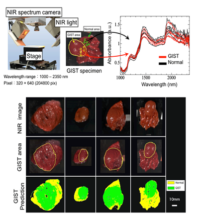
Fig.1 NIR-HSI setup and deep lesion prediction by machine learning method
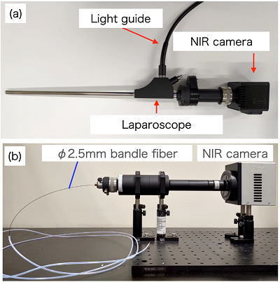
Fig.2 NIR-HSI system for laparoscope (a) and fiber scope(b)
2. Locoregional therapeutic option by laser induced plasma

Fig.3 Vaporization of tissue by laser induced plasma and future plan
Laser induced plasma can be generated by focusing pulsed laser. And when the organ is treated with the plasma, the structure of tissue is broken chemically and vaporized without impact of heat to surrounding tissue. In surgery field, it is expected as new locoregional therapeutic option which is alternative to thermal treatment such as radiofrequency ablation. Thus, we investigated the plasma vaporization effect on porcine liver in this study. As shown in Fig.3, it is observed that plasma treated area was vaporized and the surrounding tissue was free from thermal damage. We have now developed laser guide which is articulated arm type. In the future, we will develop a new device that guides the laser with fiber and can vaporize in the tissue.
3. Endoscopic Hemostasis by Low Temperature Plasma Jet

Fig.4 Low temperature plasma jet and hemostasis on porcine stomach
Recently, atmospheric Low Temperature Plasma(LTP) is focused as a novel medical tool and it is also expected to be used for hemostasis. But the size of conventional plasma source was too big to use in endoscopy, and the efficacy of LTP hemostasis was difficult to investigate for against gastrointestinal bleeding. We developed a new LTP jet collaborated with Tokyo Institute of technology, which was created with 3D printer to adapt endoscopic use(Fig. 4). Using the new device, we investigated hemostasis efficacy and safety by carbon dioxide(CO2) LTP treatment. As the result, it is confirmed that the CO2 LTP achieved hemostasis within 90 s and it was safer and gave earlier recovery.
4. Development of self-propelling endoscope

Fig.5 Developed self-propelling endoscope and insertion in colon model
Gastrointestinal endoscopy is a major diagnostic and therapeutic tool in clinical gastroenterology. However, to conduct endoscope, high level insertion technic is required. In our research, to develop automatic endoscopic diagnosis system, self-propelling device has been developing using wire and air driven method. As shown Fig.5, we found that the device can move to insertion direction by itself in gently curved colon model.