HOME > Publication & Reports > Annual Report 2016 > Hospital East
Department of Gastroenterology and Endoscopy
Tomonori Yano, Hiroaki Ikematsu, Yasuhiro Oono, Keisuke Hori, Yusuke Yoda, Tomohiro Kadota, Keiichirou Nakajyou, Kazuhiro Kaneko
Introduction
The Department of Gastroenterology and Endoscopy consists of five staff doctors who are specialized in endoscopic diagnosis and treatment for digestive tract, and head and neck cancer. The main service of our department is the endoscopic diagnosis and treatment such as endoscopic mucosal resection (EMR), and endoscopic submucosal dissection (ESD) for early stage cancers of pharyngeal region, esophagus, stomach, and colon.
Our team and what we do
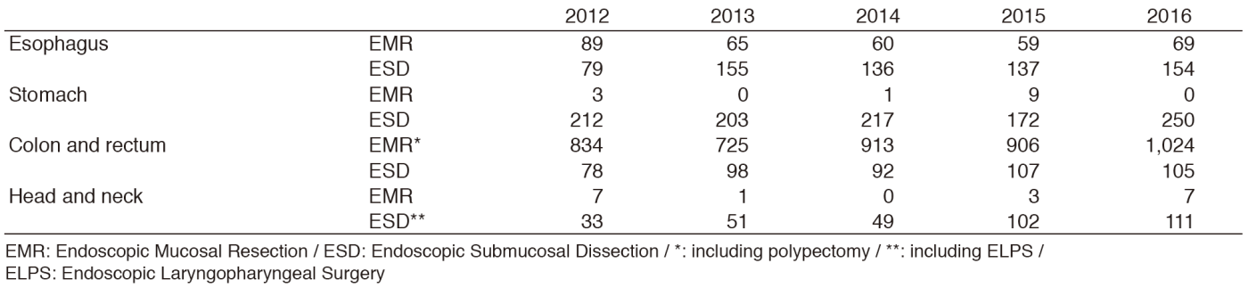
Staff members of this department performed more than a total of 12,000 endoscopic procedures, 1,700 EMR/ESD annually in 2016 (Tables 1, 2), and the number of cases are gradually increasing which ended up being the highest record ever. Furthermore, the staff members performed minimally invasive endoscopic procedure except for EMR/ESD for early stage cancer, salvage treatment such as ESD and photodynamic therapy (PDT) for local failure after radiotherapy for esophageal cancer, and palliative treatments for patients with symptomatic advanced cancer such as stent placement, percutaneous endoscopic gastroscopy, and balloon dilatation. Also, the upper GI group aggressively performed endoscopic treatment for early pharyngeal cancer through the collaboration with head and neck surgeon (Table 2). In addition, the colon group concentrated the diagnosis with fine magnified images using image enhanced endoscopy for adenoma and early cancer even in clinical practices. During the daily endoscopic examination, physicians access primary tumors through endoscopy and easily take numerous numbers of biopsy material from cancerous lesions. Recently, a lot of molecular target agents are developed and included as practices in the oncology field. Therefore, the position of endoscopy is becoming more important in clinical oncology as an easy access tool for primary tumor.
Table 1. Number of patients examined in 2012-2016
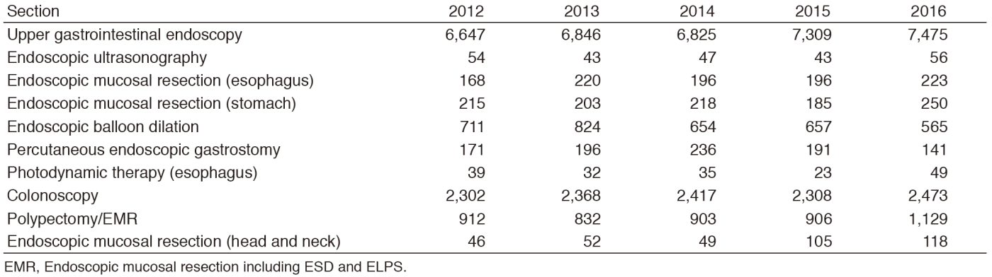
Table 2. Endoscopic procedures in 2012-2016
Research activities
The major research activities at this department are translational research, innovation for new imaging and endoscopic equipment through the collaboration with an academia or company, and clinical studies. In the field of translational research, several research works collaborate with the Department of Pathology in order to clarify the elucidation of early stage cancer in digestive tract and head and neck cancer. In 2016, papers about the natural history of pathologically confirmed superficial pharyngeal cancer and molecular characteristics of the skirt lesion around laterally spreading tumor in colon were published. Also, papers about long term survival data of patients who were treated with salvage PDT or the risk factors for local recurrence after salvage endoscopic treatment for local failure after radiotherapy for esophageal cancer were published as clinical research. Clinical trials for novel endoscopic hypoxia imaging for cancerous lesions or biodegradable stent for refractory benign stricture finished and the results of these studies were derived to a company, and became the keystone for approvals.
Clinical trials
We are participating in undergoing clinical trials to evaluate the efficacy and safety of tissue engineered cell sheets to prevent stricture after ESD for wide spreading superficial esophageal cancer. In addition, we are conducting a trial to evaluate the imaging using near-infrared light with a wavelength of over 1000nm for surgical resected GIST material. Furthermore, we are participating in many multi-institutional clinical trials including the Japan Clinical Oncology Group (JCOG) study. As for the JCOG study, phase II study of ESD for patients with undifferentiated early gastric cancer (JCOG1009/1010), phase II/III study comparing balloon dilation with steroid injection and radial incision, and the cutting method for refractory anatomical stricture after esophagectomy (JCOG1207), and phase III study comparing steroid injection and oral administration for the prevention of post esophageal ESD stricture.
Education
In our department, two senior residents and eight residents have been enrolled. Trainees could perform services under the training program with observation by each specialist. Futhermore, they are educated not only for endoscopic diagnosis and endoscopic treatment strategy, but also for pathological diagnosis, clinical or surgical oncology through the conference with other departments. In addition, they have actively presented their research works at international conferences, published papers in international peer reviewed journals, and seven papers written by residents were published in 2016. They wrote several study protocols mainly for innovative development in endoscopic imaging and treatment. For residents who want to learn more in detail about regulatory science for medical devices, they can have a chance to study in the Pharmaceuticals and Medical Device Agency (PMDA) after their graduation.
Future prospects
The new endoscopy center will open on the first floor of the Center of New Surgical and Endoscopic Development for Exploratory Technology (NEXT) in 2017. Endoscopic center will expand more than three times larger than now. The objective after opening the new endoscopy center is to increase the handling number of endoscopic diagnoses and treatments. Moreover, we want to perform innovative activities for the creation of new endoscopic images or treatment, and clinical studies in order to bring new endoscopic modalities for cancer patients, and not only now, but also for the future.
List of papers published in 2016
Journal
1.Katada C, Yokoyama T, Yano T, Kaneko K, Oda I, Shimizu Y, Doyama H, Koike T, Takizawa K, Hirao M, Okada H, Yoshii T, Konishi K, Yamanouchi T, Tsuda T, Omori T, Kobayashi N, Shimoda T, Ochiai A, Amanuma Y, Ohashi S, Matsuda T, Ishikawa H, Yokoyama A, Muto M. Alcohol Consumption and Multiple Dysplastic Lesions Increase Risk of Squamous Cell Carcinoma in the Esophagus, Head, and Neck. Gastroenterology, 151:860-869 e867, 2016
2.Uedo N, Gotoda T, Yoshinaga S, Tanuma T, Morita Y, Doyama H, Aso A, Hirasawa T, Yano T, Uchita N, Ho S-H, Hsieh P-H. Differences in routine esophagogastroduodenoscopy between Japanese and international facilities: A questionnaire survey. Dig Endosc, 28 Suppl 1:16-24, 2016
3.Sano Y, Tanaka S, Kudo S-E, Saito S, Matsuda T, Wada Y, Fujii T, Ikematsu H, Uraoka T, Kobayashi N, Nakamura H, Hotta K, Horimatsu T, Sakamoto N, Fu K-I, Tsuruta O, Kawano H, Kashida H, Takeuchi Y, Machida H, Kusaka T, Yoshida N, Hirata I, Terai T, Yamano H-O, Kaneko K, Nakajima T, Sakamoto T, Yamaguchi Y, Tamai N, Nakano N, Hayashi N, Oka S, Iwatate M, Ishikawa H, Murakami Y, Yoshida S, Saito Y. Narrow-band imaging (NBI) magnifying endoscopic classification of colorectal tumors proposed by the Japan NBI Expert Team. Dig Endosc, 28:526-533, 2016
4.Osera S, Oono Y, Ikematsu H, Yano T, Kaneko K. Endoscopic submucosal resection with a ligation device for the treatment of duodenal neuroendocrine tumors. Surg Endosc, 30:3928-3932, 2016
5.Matsuda T, Sekiguchi M, Kakugawa Y, Ikematsu H, Oono Y, Chiu H-M, Sano Y, Fujii T, Saito Y. Colorectal cancer prevention and early diagnosis using endoscopy. Nihon Shokakibyo Gakkai Zasshi, 113:1176-1185, 2016
6.Matsuda T, Oka S, Ikematsu H, Matsushita H-o, Mori Y, Takeuchi Y, Tamai N, Kawamura T, Chino A, Keum B, Khomvilai S, Uraoka T. Endoscopic diagnosis of colorectal serrated lesions: Current status and future perspectives based on the results of a questionnaire survey. Dig Endosc, 28 Suppl 1:35-42, 2016
7.Kasai M, Yasuda Y, Takemura H, Mizoguchi H, Soga K, Kaneko K. In vivo tumor spatial classification using PCA and K-means with NIR-hyperspectral data. J Biomed Eng Med Imaging, 3:45, 2016
8.Kaneko K. [Frontiers of endoscopic diagnosis for cancer -innovation of a novel endoscopy]. Nihon Shokakibyo Gakkai Zasshi, 113:205-213, 2016
9.Kadota T, Yano T, Kato T, Imajoh M, Noguchi M, Morimoto H, Osera S, Yoda Y, Oono Y, Ikematsu H, Ohtsu A, Kaneko K. Prophylactic steroid administration for strictures after endoscopic resection of large superficial esophageal squamous cell carcinoma. Endosc Int Open, 4:E1267-E1274, 2016
10.Hirasawa T, Uchita K, Yano T. How many pictures are demanded for screening gastroscopy? Dig Endosc, 28 Suppl 1:33-34, 2016
11.Gotoda T, Uedo N, Yoshinaga S, Tanuma T, Morita Y, Doyama H, Aso A, Hirasawa T, Yano T, Uchita K, Ho S-H, Hsieh P-H. Basic principles and practice of gastric cancer screening using high-definition white-light gastroscopy: Eyes can only see what the brain knows. Dig Endosc, 28 Suppl 1:2-15, 2016
12.Takizawa K, Ono H, Yamamoto Y, Katai H, Hori S, Yano T, Umegaki E, Sasaki S, Iizuka T, Kawagoe K, Shimoda T, Muto M, Sasako M. Incidence of lymph node metastasis in intramucosal gastric cancer measuring 30 mm or less, with ulceration; mixed, predominantly differentiated-type histology; and no lymphovascular invasion: a multicenter retrospective study. Gastric Cancer, 19:1144-1148, 2016
13.Kadota T, Fujii S, Oono Y, Imajoh M, Yano T, Kaneko K. Adenocarcinoma arising from heterotopic gastric mucosa in the cervical esophagus and upper thoracic esophagus: two case reports and literature review. Expert Rev Gastroenterol Hepatol, 10:405-414, 2016
14.Kakushima N, Hori K, Ono H, Horimatsu T, Uedo N, Ohata K, Doyama H, Kaneko K, Oda I, Hikichi T, Kawahara Y, Niimi K, Takaki Y, Mizuno M, Yazumi S, Hosokawa A, Imagawa A, Niimi M, Yoshimura K, Muto M. Proton pump inhibitor after endoscopic resection for esophageal squamous cell cancer: multicenter prospective randomized controlled trial. J Gastroenterol, 51:104-111, 2016
15.Osera S, Fujii S, Ikematsu H, Miyamoto H, Oono Y, Yano T, Ochiai A, Yoshino T, Ohtsu A, Kaneko K. Clinicopathological, endoscopic, and molecular characteristics of the "skirt" - a new entity of lesions at the margin of laterally spreading tumors. Endoscopy, 48:448-455, 2016
16.Hatogai K, Yano T, Kojima T, Onozawa M, Fujii S, Daiko H, Yoda Y, Hombu T, Doi T, Kaneko K, Ohtsu A. Local efficacy and survival outcome of salvage endoscopic therapy for local recurrent lesions after definitive chemoradiotherapy for esophageal cancer. Radiat Oncol, 11:31, 2016
17.Nakamura H, Yano T, Fujii S, Kadota T, Tomioka T, Shinozaki T, Hayashi R, Kaneko K. Natural history of superficial head and neck squamous cell carcinoma under scheduled follow-up endoscopic observation with narrow band imaging: retrospective cohort study. BMC Cancer, 16:743, 2016
18.Hatogai K, Yano T, Kojima T, Onozawa M, Daiko H, Nomura S, Yoda Y, Doi T, Kaneko K, Ohtsu A. Salvage photodynamic therapy for local failure after chemoradiotherapy for esophageal squamous cell carcinoma. Gastrointest Endosc, 83:1130-1139 e1133, 2016