HOME > Publication & Reports > Annual Report 2016 > Hospital
Department of Diagnostic Radiology (Interventional Radiology Center)
Yasuaki Arai, Yasunori Mizuguchi, Gen Iinuma, Miyuki Sone, Hiroaki Kurihara, Nachiko Uchiyama, Minoru Machida, Mari Kikuchi, Hirokazu Watanabe, Tomoko Manabe, Eriko Iwamoto, Mototaka Miyake, Hideaki Kobayashi, Syunsuke Sugawara, Yuko Kubo, Takahiro Morita, Koji Tomita, Sayako Iwashita, Shinji Wada, Takatoshi Kubo, Keishi Fujiwara
Introduction
The Department of Diagnostic Radiology provides a wide range of modalities, including interventional radiology (IVR), general radiology, computed tomography (CT), magnetic resonance imaging (MRI), ultrasound, mammography and nuclear medicine.
In 2014, we launched the Center for Interventional Radiology to facilitate widespread proliferation of IVR in Japan and to provide various IVR treatments for the patients referred from other hospitals and clinics.
We seek individuals with outstanding leadership capabilities, proven academic and administrative experience, the vision to build and sustain programs at the forefront of imaging research, and a commitment to clinical experience.
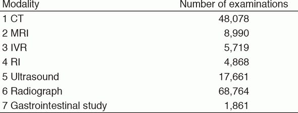
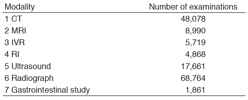
Table 1. Routine activities
Research activities
The CT colonography (CTC) has been successfully introduced as an effective option for preoperative staging and colorectal screening in our center. Nearly 1,000 patients and/or candidates were examined with this modality in 2016. For the preparation of screening CTC, electronic cleansing with fecal barium tagging and automated CO2 gas insufflation systems have been established in the formal National Cancer Center (NCC) collaboration studies with the associated companies. Furthermore, we are now developing computer-aided detection (CAD) for colorectal lesions, especially for flat lesions. The main purpose of our CTC research work is to conduct a multicenter trial to establish evidence regarding fully digitalized CTC for a colorectal screening system in Japan.
A multi tracer consisting of [18F]-FDG, [18F]-FBPA, [11C]-choline, [11C]-methionine, and [64Cu]-DOTA-antibody PET imaging has been studied for cancer patients to improve the sensitivity and specificity of detecting tumor sites or tumor characteristics. [18F]-FDG dynamic PET sampling with Patrak-plot analysis allows us to calculate the glucose metabolic rate of the tumor site. [18F]-FBPA PET/CT has been conducted in over 100 cancer patients through the clinical research. [11C]-choline and [11C]-methionine PET/CT examinations have been scheduled routinely two days per week. As for [64Cu]-DOTA-antibody PET imaging, [64Cu]-DOTA-trastuzumab PET/CT has been conducted in HER-2 positive breast cancer patients. Respiratory-gated PET/CT was evaluated to reduce breathing-induced artifacts using a four-dimensional PET/CT protocol. It provided better localization and quantification of tumors around the lower thorax to the upper abdomen. A newly installed scanner of PET/MRI has been applied to more than 700 cases of cancer patients and proved itself to be a powerful tool for managing malignancy. For cancer treatment, internal radiotherapy was carried out with the use of radioactive iodine (I-131) chloride, Zevalin, metastron, and Xofigo.
In accordance with the achievement of collaborative research with the associated company since 2009, digital breast tomosynthesis (DBT) has been introduced as an effective routine option for preoperative evaluation since March 2014. Up to December 2016, 1,835 patients were examined. Regarding image acquisition systems, new software-based scatter correction enabling gridless has been developed and acquired in clinical usage. In addition, new volumetric image reconstruction algorisms such as synthetic 2D, synthetic 3D, and thick slab images have been developed and evaluated to decrease radiation dose and reading number of images.
A multicenter study to establish the CT classification of lung adenocarcinomas corresponding to the new International Association for the Study of Lung Cancer, American Thoracic Society and European Respiratory Society (IASLC/ATS/ERS) pathological classification and to build the database of small adenocarcinomas has been performed. The Digital Imaging and Communications in Medicine (DICOM) data of resected lung cancers from each institute have been accumulated and evaluated in collaboration with the Japanese Society of Thoracic Radiology.
The Japan Response Evaluation Criteria in Solid Tumors (RECIST) working group has developed a tumor response evaluation computer system, which is capable of semiautomatic RECIST evaluation and is compliant with DICOM data.
Image guided preoperative Breast Marking using ultrasound alone or combined with mammography has been performed for partial mastectomy cases which are difficult to determine the spread of disease. This technique makes it possible to resect abnormal lesion more precisely and assists to prevent both re-operation and local recurrence. A total of 47 cases were performed from January to December 2016.
Clinical trials
A major departmental research theme is establishing an evidence base for interventional radiology (IVR). We have led a multiinstitutional cooperative study group of IVR (JIVROSG: Japan Interventional Radiology in Oncology Study Group) since 2002 as a steering organization of 90 participating domestic institutions. In this study group, we investigate the efficacy of palliative interventional radiology in randomized controlled trials (RCTs) to compare it with other therapies. These palliative RCTs include: a phase III study evaluating the efficacy of peritoneo-venous shunting (JIVROSG-0803); a phase III study evaluating the efficacy of percutaneous vertebroplasty for painful bone metastases (JIVROSG-0804); a phase III study evaluating the efficacy of percutaneous trans-esophageal gastric tubing (JIVROSG-0805); a phase III study evaluating the efficacy of stenting for SVC/IVC syndrome (JIVROSG-0807), and JIVROSG-0807 completed patient enrollment in 2013. Other ongoing clinical trials are a phase II study evaluating the efficacy of arterial infusion chemotherapy and radiotherapy for unresectable maxillary carcinoma (JIVROSG-0808), a phase II trial of palliative intra-arterial epirubicin/5FU therapy for patients with chemotherapy-refractory locally-advanced or metastatic breast cancer (JIVROSG-1107), and a prospective randomized controlled trial of selective DEB-epiDOX vs. selective conventional TACE for hepatocellular carcinoma focusing on local complete response rate (JIVROSG-1302: PRESIDENR study).
Education
The clinical education and training of young radiologists is an important part of our department's activities. During 2016, eight residents and four short-term residents were trained in our department. Educational opportunities for three foreign physicians from U.S.A., China, and Singapore, were also provided. We hold several clinical or educational conferences. A daily clinical IVR case conference, a weekly educational case conference on diagnostic radiology, and a monthly IVR research conference are held.
Future prospects
The Department of Diagnostic Radiology aims to strive for excellence in clinical care, education, and research. Our goal is to provide outstanding patient-centered radiology services and to establish evidence in this area. Future challenges include promoting the active role of the Center for Interventional Radiology, which opened last year, and facilitating imaging as biomarkers for personalized cancer treatments such as molecular-targeted agents, immunotherapy, and boron neutron capture therapy.
List of papers published in 2016
Journal
1.Kurihara H, Shimizu C, Miyakita Y, Yoshida M, Hamada A, Kanayama Y, Yonemori K, Hashimoto J, Tani H, Kodaira M, Yunokawa M, Yamamoto H, Watanabe Y, Fujiwara Y, Tamura K. Molecular imaging using PET for breast cancer. Breast Cancer, 23:24-32, 2016
2.Kubo T, Kiryu S, Akai H, Ota Y, Tojo A, Yoshida H, Kato N, Nakano Y, Ohtomo K. Hepatic Involvement of Histiocytic Sarcoma: CT and MRI Findings. Korean J Radiol, 17:758-762, 2016
3.Tomita K, Hiraki T, Gobara H, Fujiwara H, Iguchi T, Matsui Y, Kanazawa S. Evaluation of Lung Radiofrequency Ablation With Dual-Energy Computed Tomography: Analysis of Tumor Composition and Lung Perfusion. J Comput Assist Tomogr, 40:752-756, 2016
4.Tsushima Y, Ishiguchi T, Murakami T, Hayashi H, Hayakawa K, Fukuda K, Korogi Y, Sugimoto H, Takehara Y, Narumi Y, Arai Y, Kuwatsuru R, Yoshimitsu K, Awai K, Kanematsu M, Takagi R. Safe use of iodinated and gadolinium-based contrast media in current practice in Japan: a questionnaire survey. Jpn J Radiol, 34:130-139, 2016
5.Tozaki M, Kuroki Y, Kikuchi M, Kojima Y, Kubota K, Nakahara H, Ito Y, Mukai H. The Japanese Breast Cancer Society clinical practice guidelines for screening and imaging diagnosis of breast cancer, 2015 edition. Breast Cancer, 23:357-366, 2016
6.Tanaka M, Kishi Y, Esaki M, Nara S, Miyake M, Hiraoka N, Nagino M, Shimada K. Feasibility of Routine Application of Gadoxetic Acid-Enhanced MRI in Combination with Diffusion-Weighted MRI for the Preoperative Evaluation of Colorectal Liver Metastases. Ann Surg Oncol, 23:3991-3998, 2016
7.Hori M, Onaya H, Hiraoka N, Yamaji T, Kobayashi H, Takahashi M, Mutoh M, Shimada K, Nakagama H. Evaluation of the degree of pancreatic fatty infiltration by area-based assessment of CT images: comparison with histopathology-based and CT attenuation index-based assessments. Jpn J Radiol, 34:667-676, 2016
8.Gobara H, Arai Y, Kobayashi T, Yamakado K, Inaba Y, Kodama Y, Yamagami T, Sone M, Watanabe H, Okumura Y, Shinya T, Kurihara H, Kanazawa S. Percutaneous radiofrequency ablation for patients with malignant lung tumors: a phase II prospective multicenter study (JIVROSG-0702). Jpn J Radiol, 34:556-563, 2016
9.Ikeda S, Manabe T, Sugawara S, Sone M, Ishikawa M, Kato T. Spontaneous rupture of a deep femoral pseudoaneurysm mimicking lymphedema after radical hysterectomy in a woman who was receiving warfarin. J Med Cases, 7:299-302, 2016
10.Kobayashi K, Kurihara H, Watanabe Y, Murakami N, Inaba K, Nakamura S, Wakita A, Okamoto H, Umezawa R, Takahashi K, Igaki H, Ito Y, Yoshimoto S, Shigematsu N, Itami J. In vivo spatial correlation between 18F-BPA and 18F-FDG uptakes in head and neck cancer. Appl Radiat Isot, 115:138-146, 2016
11.de Baere T, Arai Y, Lencioni R, Geschwind JF, Rilling W, Salem R, Matsui O, Soulen MC. Treatment of Liver Tumors with Lipiodol TACE: Technical Recommendations from Experts Opinion. Cardiovasc Intervent Radiol, 39:334-343, 2016
12.Tamura A, Kato K, Suzuki M, Sone M, Tanaka R, Nakasato T, Ehara S. CT-Guided Percutaneous Radiologic Gastrostomy for Patients with Head and Neck Cancer: A Retrospective Evaluation in 177 Patients. Cardiovasc Intervent Radiol, 39:271-278, 2016
13.Mimura H, Arai Y, Yamakado K, Sone M, Takeuchi Y, Miki T, Gobara H, Sakuhara Y, Yamamoto T, Sato Y, Kanazawa S. Phase I/II Study of Radiofrequency Ablation for Malignant Renal Tumors: Japan Interventional Radiology in Oncology Study Group 0701. Cardiovasc Intervent Radiol, 39:717-723, 2016
14.Miyazaki M, Arai Y, Myoui A, Gobara H, Sone M, Rosenthal DI, Tsushima Y, Kanazawa S, Ehara S, Endo K. Phase I/II Multi-Institutional Study of Percutaneous Radiofrequency Ablation for Painful Osteoid Osteoma (JIVROSG-0704). Cardiovasc Intervent Radiol, 39:1464-1470, 2016
15.Osuga K, Nakajima Y, Sone M, Arai Y, Nambu Y, Hori S. Transarterial embolization of hypervascular tumors using trisacryl gelatin microspheres (Embosphere): a prospective multicenter clinical trial in Japan. Jpn J Radiol, 34:366-375, 2016
16.Setsu N, Miyake M, Wakai S, Nakatani F, Kobayashi E, Chuman H, Hiraoka N, Kawai A, Yoshida A. Primary Retroperitoneal Myxoid Liposarcomas. Am J Surg Pathol, 40:1286-1290, 2016
17.Sofue K, Takeuchi Y, Tsurusaki M, Shibamoto K, Sakamoto N, Kitajima K, Sone M, Sugimura K, Arai Y. Value of Percutaneous Radiologic Gastrostomy for Patients with Advanced Esophageal Cancer. Ann Surg Oncol, 23:3623-3631, 2016
18.Sone M, Arai Y, Sugawara S, Tomita K, Fujiwara K, Ishii H, Morita S. Angio-CT-Assisted Balloon Dissection: Protection of the Adjacent Intestine during Cryoablation for Patients with Renal Cancer. J Vasc Interv Radiol, 27:1414-1419, 2016
19.Tokuda T, Arai Y, Sone M, Sugawara S, Morita S, Saito Y. Coil Embolization for the Treatment of Esophageal Perforation after Endoscopic Submucosal Dissection. J Vasc Interv Radiol, 27:1461-1463, 2016
Book
1.Nachiko Uchiyama, Mari Kikuchi, Minoru Machida,et.al. Diagnostic Usefulness of Synthetic MMG (SMMG) with DBT (Digital Breast Tomosynthesis) for Clinical Setting in Breast Cancer Screening. In: Breast Imaging, Switzerland, Springer International Publishing AG, pp 59-67, 2016