Annual Report 2017
Division of Science and Technology for Endoscopy (Kashiwa Campus)
Hiroaki Ikematsu, Tomonori Yano, Keisuke Hori, Toshihiro Takamatsu, Takako Mamada
Introduction
Now, endoscopy is widely used for screening, diagnosis, and treatment for early cancer in aero-digestive tracts including the pharynx, esophagus, stomach, and colorectum. With conventional endoscopy, observations are made using white light to illuminate the mucosal surface with special attention paid to the appearance of reddish and irregular portions compared to adjacent areas. Thus, the detection of suspicious early cancerous lesions has been largely based on the macroscopic characteristics of the lesions. Still, the detection is difficult for all endoscopists. Therefore, we are working on the development of auxiliary diagnosis for the detection of gastrointestinal lesions by using artificial intelligence (AI).
Another characteristic of a tumor is hypoxia. As a tumor grows, it rapidly outgrows its blood supply, leaving portions of the tumor with regions where the oxygen concentration is significantly lower than in healthy tissues. Thus, there have been attempts to visualize spatial distribution of tumor hypoxia, such as fluorescent labeling techniques or hemoglobin absorption-based techniques. However, these methods are limited because of low spatiotemporal resolution. We developed an imaging technology that can derive oxygen saturation (StO2) images from small numbers of wavelength measurements.
The next-generation novel endoscopy will be required to make visible specific functions in cancerous tissues.
Research activities
Present research activities mainly focus on the development of new endoscopic imaging modalities.
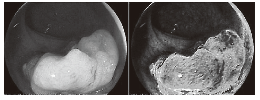
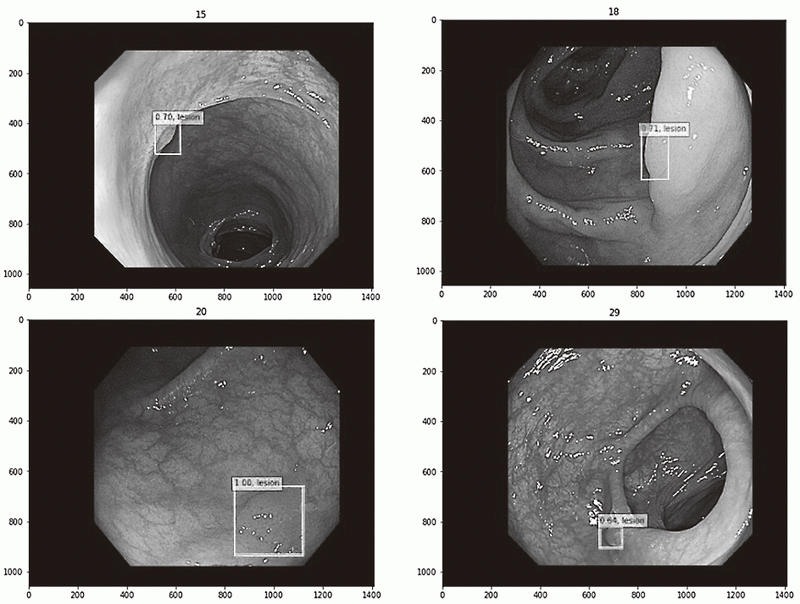
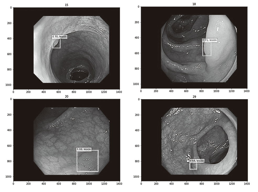
Oxygen saturation imaging is detected for neoplastic lesions of the head and neck and alimentary tracts, with two types of visualized images: a pseudocolor StO2 image and an StO2 overlay image (Figure 1). Another project is the development of diagnostic artificial intelligence (AI) system for the detection of gastrointestinal lesions with high efficiency of deep learning. Within this period, we succeeded in developing the AI model for the detection of colon lesions with high accuracy rate (Figure 2). Another project is a new bioimaging system using near-infrared light with a wavelength of over 1,000 nm with various spectrums. This system is capable of penetrating through the gastrointestinal wall and obtaining images.
Figure 1. Oxygen saturation imaging of rectal cancer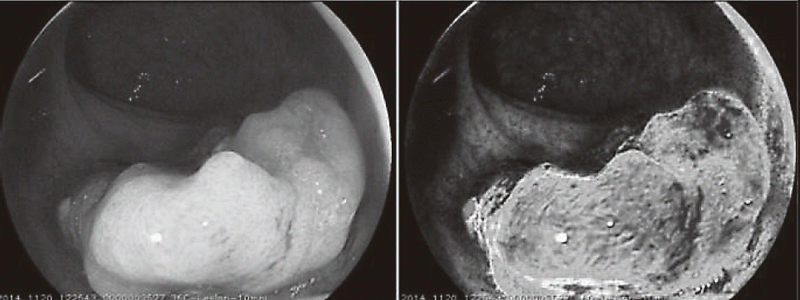
Clinical trials
1) POC study of SO2 imaging endoscopy (UMIN 000008402)
2) Development of diagnostic artificial intelligence (AI) system for the detection of gastrointestinal lesions
3) Development of diagnostic endoscopy for GIST using near-infrared light
Education
Meetings are constantly conducted with faculties including technology and science students of the university.
Future prospects
Researchers at Tokyo University of Science are scheduled to be sent to our hospital (National Cancer Center Hospital East : NCCHE) by the cross-appointment with Tokyo University of Science.
In the future, we will continue to develop endoscopic equipment in cooperation with the Department of Gastroenterology and Endoscopy of the NCCHE and try innovative approaches to produce all-new endoscopy in collaboration with academia and companies.
List of papers published in January 2017 - March 2018
Journal
1. Sunagawa H, Kinoshita T, Kaito A, Shibasaki H, Kaneko K, Ochiai A, Ohtsu A, Nishida T. Additional surgery for non-curative resection after endoscopic submucosal dissection for gastric cancer: a retrospective analysis of 200 cases. Surg Today, 47:202-209, 2017
2. Morimoto H, Yano T, Yoda Y, Oono Y, Ikematsu H, Hayashi R, Ohtsu A, Kaneko K. Clinical impact of surveillance for head and neck cancer in patients with esophageal squamous cell carcinoma. World journal of gastroenterology, 23:1051-1058, 2017
3. Kadota T, Yano T, Fujita T, Daiko H, Fujii S. Submucosal Invasive Depth Predicts Lymph Node Metastasis and Poor Prognosis in Submucosal Invasive Esophageal Squamous Cell Carcinoma. Am J Clin Pathol, 148:416-426, 2017
4. Nakanishi H, Doyama H, Ishikawa H, Uedo N, Gotoda T, Kato M, Nagao S, Nagami Y, Aoyagi H, Imagawa A, Kodaira J, Mitsui S, Kobayashi N, Muto M, Takatori H, Abe T, Tsujii M, Watari J, Ishiyama S, Oda I, Ono H, Kaneko K, Yokoi C, Ueo T, Uchita K, Matsumoto K, Kanesaka T, Morita Y, Katsuki S, Nishikawa J, Inamura K, Kinjo T, Yamamoto K, Yoshimura D, Araki H, Kashida H, Hosokawa A, Mori H, Yamashita H, Motohashi O, Kobayashi K, Hirayama M, Kobayashi H, Endo M, Yamano H, Murakami K, Koike T, Hirasawa K, Miyaoka Y, Hamamoto H, Hikichi T, Hanabata N, Shimoda R, Hori S, Sato T, Kodashima S, Okada H, Mannami T, Yamamoto S, Niwa Y, Yashima K, Tanabe S, Satoh H, Sasaki F, Yamazato T, Ikeda Y, Nishisaki H, Nakagawa M, Matsuda A, Tamura F, Nishiyama H, Arita K, Kawasaki K, Hoppo K, Oka M, Ishihara S, Mukasa M, Minamino H, Yao K. Evaluation of an e-learning system for diagnosis of gastric lesions using magnifying narrow-band imaging: a multicenter randomized controlled study. Endoscopy, 49:957-967, 2017
5. Nakamura H, Ikematsu H, Osera S, Ito R, Sato D, Minamide T, Okamoto N, Yamamoto Y, Hombu T, Takashima K, Nakajo K, Kadota T, Yoda Y, Hori K, Oono Y, Yano T. Visual assessment of colorectal flat and depressed lesions by using narrow band imaging. Endoscopy international open, 5:E1284-E1288, 2017
6. Yokoyama A, Katada C, Yokoyama T, Yano T, Kaneko K, Oda I, Shimizu Y, Doyama H, Koike T, Takizawa K, Hirao M, Okada H, Yoshii T, Konishi K, Yamanouchi T, Tsuda T, Omori T, Kobayashi N, Suzuki H, Tanabe S, Hori K, Nakayama N, Kawakubo H, Ishikawa H, Muto M. Alcohol abstinence and risk assessment for second esophageal cancer in Japanese men after mucosectomy for early esophageal cancer. PLoS One, 12:e0175182, 2017
7. Oka S, Uraoka T, Tamai N, Ikematsu H, Chino A, Okamoto K, Takeuchi Y, Imai K, Ohata K, Shiga H, Raftopoulos S, Lee BI, Matsuda T. Standardization of endoscopic resection for colorectal tumors larger than 10 mm in diameter. Dig Endosc, 29 Suppl 2:40-44, 2017
8. Hotta K, Matsuda T, Kakugawa Y, Ikematsu H, Kobayashi N, Kushima R, Hozawa A, Nakajima T, Sakamoto T, Mori M, Fujii T, Saito Y. Regional colorectal cancer screening program using colonoscopy on an island: a prospective Nii-jima study. Jpn J Clin Oncol, 47:118-122, 2017
9. Ikematsu H, Sakamoto T, Togashi K, Yoshida N, Hisabe T, Kiriyama S, Matsuda K, Hayashi Y, Matsuda T, Osera S, Kaneko K, Utano K, Naito Y, Ishihara H, Kato M, Yoshimura K, Ishikawa H, Yamamoto H, Saito Y. Detectability of colorectal neoplastic lesions using a novel endoscopic system with blue laser imaging: a multicenter randomized controlled trial. Gastrointest Endosc, 86:386-394, 2017
10. Niwa A, Kuwano S, Tomita H, Kimura K, Orihara Y, Kanayama T, Noguchi K, Hisamatsu K, Nakashima T, Hatano Y, Hirata A, Miyazaki T, Kaneko K, Tanaka T, Hara A. The different pathogeneses of sporadic adenoma and adenocarcinoma in non-ampullary lesions of the proximal and distal duodenum. Oncotarget, 8:41078-41090, 2017
11. Yano T, Yoda Y, Nomura S, Toyosaki K, Hasegawa H, Ono H, Tanaka M, Morimoto H, Horimatsu T, Nonaka S, Kaneko K, Sato A. Prospective trial of biodegradable stents for refractory benign esophageal strictures after curative treatment of esophageal cancer. Gastrointest Endosc, 86:492-499, 2017
12. Osera S, Ikematsu H, Fujii S, Hori K, Oono Y, Yano T, Kaneko K. Endoscopic treatment outcomes of laterally spreading tumors with a skirt (with video). Gastrointest Endosc, 86:533-541, 2017
13. Noguchi M, Yano T, Kato T, Kadota T, Imajoh M, Morimoto H, Osera S, Yagishita A, Odagaki T, Yoda Y, Oono Y, Ikematsu H, Kaneko K. Risk factors for intraoperative perforation during endoscopic submucosal dissection of superficial esophageal squamous cell carcinoma. World journal of gastroenterology, 23:478-485, 2017
14. Yano T, Kasai H, Horimatsu T, Yoshimura K, Teramukai S, Morita S, Tada H, Yamamoto Y, Kataoka H, Kakushima N, Ishihara R, Isomoto H, Muto M. A multicenter phase II study of salvage photodynamic therapy using talaporfin sodium (ME2906) and a diode laser (PNL6405EPG) for local failure after chemoradiotherapy or radiotherapy for esophageal cancer. Oncotarget, 8:22135-22144, 2017
15. Kanesaka T, Uedo N, Yao K, Ezoe Y, Doyama H, Oda I, Kaneko K, Kawahara Y, Yokoi C, Sugiura Y, Ishikawa H, Takeuchi Y, Arao M, Iwatsubo T, Iwagami H, Matsuno K, Muto M, Saito Y, Tomita Y. Multiple convex demarcation line for prediction of benign depressed gastric lesions in magnifying narrow-band imaging. Endosc Int Open, 6:E145-E155, 2018
16. Niwa A, Kuwano S, Tomita H, Kimura K, Orihara Y, Kanayama T, Noguchi K, Hisamatsu K, Nakashima T, Hatano Y, Hirata A, Miyazaki T, Kaneko K, Tanaka T, Hara A. Correction: The different pathogenesis of sporadic adenoma and adenocarcinoma in non-ampullary lesions of the proximal and distal duodenum. Oncotarget, 9:11427, 2018
17. Hasuike N, Ono H, Boku N, Mizusawa J, Takizawa K, Fukuda H, Oda I, Doyama H, Kaneko K, Hori S, Iishi H, Kurokawa Y, Muto M. A non-randomized confirmatory trial of an expanded indication for endoscopic submucosal dissection for intestinal-type gastric cancer (cT1a): the Japan Clinical Oncology Group study (JCOG0607). Gastric Cancer, 21:114-123, 2018
18. Murakami H, Horiba R, Iwata T, Miki Y, Uno B, Sakai T, Kaneko K, Ishihama Y, Teshima N, Esaka Y. Progress in a selective method for the determination of the acetaldehyde-derived DNA adducts by using HILIC-ESI-MS/MS. Talanta, 177:12-17, 2018