Annual Report 2017
Department of Radiological Technology
Yoshihisa Muramatsu, Shoichi Katsuta, Kazunori Yokoyama, Kazuyoshi Yamano, Yuichi Nagai, Koichi Nemoto, Masashi Iseya, Takaki Ariji, Kuniji Naoi, Naotaka Yamazawa, Satoe Kitoh, Tsunemichi Akita, Hajime Ohyoshi, Kaoru Ikeno, Hiroyuki Ohta, Yukie Ishikawa, Hiroshi Tsuruoka, Keiichi Nomura, Daiki Kumagai, Fuminori Shimizu, Asami Tanaka, Ryuzo Uehara, Syogo Amano, Kaori Yanagisawa, Yukihiro Matsukawa, Toshiyuki Shibuya, Kazuya Mizuguchi, Yuto Iwabuchi, Yuki Tanaka, Syun Aoyagi, Kieji Yoshihara, Mitsuru Godo, Hikaru Kugahara, Hiroyuki Asai, Kazuto Kano, Toshiya Rachi, Kouta Hirotaki, Daiki Kanke, Taku Tochinai, Yohei Takeda, Akiho Mori, Naoki Yamashita, Mayu Wakabayashi, Chikara Mano, Mieka Funakoshi, Hikari Inagawa, Hiroaki Sagara, Takashi Someya, Yumika Hirai, Ryutaro Kawamura
Routine activities and research activities
The number of radiological examinations continues to increase in recent years (Table 1). The X-ray fluoroscopic examinations were consolidated within the Center of New Surgical and Endoscopic Development for Exploratory Technology (NEXT). The barium enema shifted to the CT colonography. A mammography imaging device was updated to a model with tomosynthesis function and we worked on the evaluation of detailed form and continuity of the lesion. For photon beam therapy, we actively promoted image-guided radiation therapy in addition to intensity-modulated radiotherapy and stereotactic body radiotherapy. For proton beam therapy, irradiation by line scanning method which performed under advanced control was applied to prostate cancer. Past X-ray films were discarded leaving a part of permanent preservation, and the storage in the film-house came to an end.
Table 1. Transition of number of radiological examination and radiation therapy by year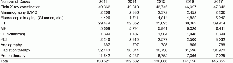
Research findings
We got a grant for overseas trainees and dispatched technologists to Duke University Hospital for on-site training of PET / CT examinations and to Charing Cross Hospital for quality control training of mammography. As joint research with companies, we reached the first contract as the Department of Radiological Technology alone. It is the development of a software to support radiation work in general, and continuous activities are expected. Regarding photon beam therapy, we reported about skin dose estimation and about evaluation of dose distribution based on different calculation algorithms. In the proton beam therapy system, we participated in the development of technology for Intensity - modulated proton therapy which administrates dose to the limited region.
Education
From this fiscal year, we have newly opened the National Cancer Center Hospital East (NCCHE) Radiological Technology Conference. Its frequency is about twice a year and each meeting consists of two parts. One part is a lecture by an invited speaker to get information related to the latest medical technology, and another part is a presentation from our hospital about cancer diagnosis and therapeutic technique, collaborating with surrounding hospitals. The result of training on human resources is as follows: the first semester of Ph.D. (master's degree) course; one person, screening examination of chief technologists in NHO; three persons passed, technologist expert in lung cancer CT screening; one person, technologist expert in breast cancer screening; one person, radiotherapy technologist; one person, radiation therapy quality manager; one person, and health officer's license on industrial hygiene; one person. In addition, we accepted nine clinical interns from four training schools for medical radiology technologists and we dispatched lecturers to three training schools.
Future prospects
In the next fiscal year, a large-scale structural change of the radiology department, a full version upgrade of MRI equipment, and the renewal of linear accelerator are planned in the radiotherapy department, respectively. We are going to proceed with renovations under good preparation to minimize the influence on routine practice.
List of papers published in January 2017 - March 2018
Journal
1. Yamazaki D, Miyazaki O, Takei Y, Matsubara K, Shinozaki M, Shimada Y, Suzuki S, Muramatsu Y. USEFULNESS OF SIZE-SPECIFIC DOSE ESTIMATES IN PEDIATRIC COMPUTED TOMOGRAPHY: REVALIDATION OF LARGE-SCALE PEDIATRIC CT DOSE SURVEY DATA IN JAPAN. Radiat Prot Dosimetry, 1-9, 2017
2. Hosokawa S, Inoue K, Kano D, Shimizu F, Koyama K, Nakagami Y, Muramatsu Y, Fukushi M. A simulation study for estimating scatter fraction in whole-body 18F-FDG PET/CT. Radiol Phys Technol, 10:204-212, 2017
3. Fujii K, Nomura K, Muramatsu Y, Obara S, Akahane K, Kusumoto M. Organ Dose Evaluations Based on Monte Carlo Simulation for CT Examinations Using Tube Current Modulation. Radiat Prot Dosimetry, 174:387-394, 2017
4. Rachi T, Hotta K, Baba H. The verification of the dose calculation accuracy of Simplified Monte Carlo method on proton beam therapy. Proceedings of the 114th Scientific Meeting of JSMP, 37:160, 2017