Annual Report 2017
Department of Endoscopy, Respiratory Endoscopy Division (Endoscopy Center)
Takaaki Tsuchida, Yuji Matsumoto, Midori Tanaka
Introduction
For respiratory diseases, we have focused on the accurate and less invasive diagnosis of miniature peripheral malignancies detected by computed tomography (CT), which can lead to early surgical treatment and less-invasive treatment including bronchoscopic therapies. This is facilitated by a multi-purpose bronchoscopy system consisting of a flat-panel fluoroscope, as well as with patient cooperation and appropriate support by medical personnel.
Endobronchial malignancies are diagnosed with videobronchoscopy, together with an endobronchial ultrasound system, and a high-
resolution flat-panel fluoroscope. In addition, imaging diagnosis, including that with high-
resolution CT, is also a routine activity for bronchoscopy, which leads to more accurate and safer diagnoses and the earlier detection of tracheobronchial malignancies.
Our team and what we do
Endobronchial ultrasonography (EBUS) is used not only to evaluate mediastinal or hilar malignant lesions, but also to evaluate whether biopsy devices can be directed to peripheral lung lesions. Two hundred eighty-five cases of EBUS-TBNA (EBUS-trans bronchial needle aspiration) were performed as a less invasive procedure to improve diagnosis for patients with mediastinal or hilar lymph node swelling. The EBUS-GS (guide sheath) method with virtual navigation was performed in most of peripheral pulmonary lesions.
Endobronchial stenosis patients were treated with airway stent placement, photodynamic therapy and/or endobronchial electrocautery ablation. Medical thoracoscopy under local anesthesia in the endoscopic examination room was performed for unknown pleural effusion or pleural tumors.
Research activities
Our efforts have been focused on new diagnostic and therapeutic strategies including bronchoscopy, which involve CT-screening for lung cancer and lead to cure and less-invasive treatments for lung cancer. To achieve more accurate endoscopic diagnosis for solitary peripheral lung nodules, we are using three-dimensional computed tomography (3D-CT) navigation, ultrasound-guided approach, electromagnetic navigation, and/or on-site cytology.
Table 1. Type of procedure and number of patients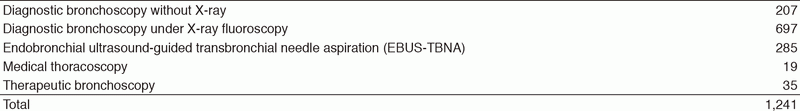
We showed the utility of rapid on-site cytologic evaluation during EBUS-TBNA. We also showed the utility of bronchoscopic procedure for re-biopsy.
Clinical trials
We conducted a multicenter prospective study for evaluation of photodynamic therapy for peripheral lung cancer.
A multicenter study for the evaluation of virtual bronchoscopic navigation with CT workstation was also performed.
Education
A flexible bronchoscope was developed for the first time in the world in the National Cancer Center Hospital (NCCH). There are many residents and overseas doctors wishing to train their skill at our hospital. We gave many residents opportunities for writing papers and delivering conference presentations. Overseas doctors from many countries trained at our hospital, making the most of what they have learned here, are active in their own country.
Future prospects
The development of a new diagnostic method with a compact electron microscope and a multicenter clinical trial for the evaluation of photodynamic therapy for peripheral lung cancer are expected to be carried out.
List of papers published in January 2017 - March 2018
Journal
1. Okubo Y, Matsumoto Y, Nakai T, Tsuchida T, Asakura K, Motoi N, Watanabe SI. The new transbronchial diagnostic approach for the metastatic lung tumor from renal cell carcinoma-a case report. J Thorac Dis, 9:E762-E766, 2017
2. Izumo T, Matsumoto Y, Sasada S, Chavez C, Nakai T, Tsuchida T. Utility of rapid on-site cytologic evaluation during endobronchial ultrasound with a guide sheath for peripheral pulmonary lesions. Jpn J Clin Oncol, 47:221-225, 2017
3. Nakai T, Matsumoto Y, Suzuk F, Tsuchida T, Izumo T. Predictive factors for a successful diagnostic bronchoscopy of ground-glass nodules. Ann Thorac Med, 12:171-176, 2017
4. Nakai T, Izumo T, Matsumoto Y, Tsuchida T. Virtual fluoroscopy during transbronchial biopsy for locating ground-glass nodules not visible on X-ray fluoroscopy. J Thorac Dis, 9:5493-5502, 2017
5. Matsumoto Y, Izumo T, Sasada S, Tsuchida T, Ohe Y. Diagnostic utility of endobronchial ultrasound with a guide sheath under the computed tomography workstation (ziostation) for small peripheral pulmonary lesions. Clin Respir J, 11:185-192, 2017