Annual Report 2018
Department of Diagnostic Radiology
Tatsushi Kobayashi, Hirofumi Kuno, Yasunori Arai, Kaoru Shimada, Takashi Hiyama, Shioto Oda
Introduction
The Department of Diagnostic Radiology is committed to improving health through excellence in image-oriented patient care and research. Our department performs approximately 100,000 inpatient and outpatient procedures annually. Our department also conducts clinical scientific research as well as basic scientific studies, with the results translated directly into better patient care.
The Team and What We Do
Our department has four multi-slice CT scanners including two area detector CT scanners and one Dual Source CT, two 3T MRI systems, one interventional radiology (IR) CT system, one Multi-axis c-arm CT system, two gamma cameras with the capacity for single photon emission CT (SPECT), two digital radiographic (DR) systems for fluoroscopy, two mammographies (MMG), and four computed radiographic (CR) systems. Our IR-CT systems use digital subtraction angiography with multi-detector computerized tomography (MDCT). One is equipped with a 320 multi-slice CT. A positron emission tomography (PET) scanner and baby cyclotron have been installed, and tumor imaging using 18F-FDG (fluorodeoxyglucose) has been performed. These all-digital image systems enhance the efficacy of routine examinations.
This department has eight consulting radiologists and 22 technologists. As part of our routine activities, every effort is made to produce an integrated report covering almost all examinations, such as MMG, contrast radiological procedures, CT, MRI, RI, PET, angiography and IR, mainly transarterial chemoembolization (TACE).
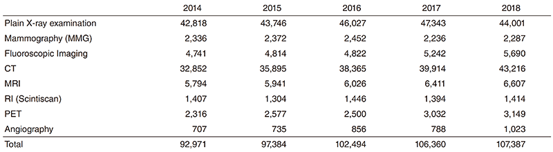
The number of cases examined in 2018 is shown in Table 1. Several conferences are routinely held at our department including preand postoperative conferences. Furthermore, our department contributes to decide the treatment strategy through the image presentation at the weekly tumor board conference (especially, Hepatobiliary-Pancreatic and Head-Neck regions).
Table 1. Number of examinations
Research activities
The research activities of the Department of Diagnostic Radiology focus on diagnostic imaging and IR. These activities consist of: 1) Development of new CT/MR Imaging techniques and 2) Texture analysis and radiomics research. Our department also conducts clinical scientific research as well as basic scientific studies, with the results translated directly into better patient care.
1. Development of new CT/MR Imaging techniques
A major focus of our department is in the development of new imaging techniques using advanced CT/MR systems including dual-energy CT (DECT) and area-detector CT (ADCT) for cancer patients.
DECT has the potential to improve detection of pathologies and increase diagnostic confidence in the evaluation of a variety of cancers by using different X-ray energy-dependent absorption behaviors of different materials. DECT allows material decomposition so that iodine can be differentiated from soft tissue, and can potentially provide additional further "contrast resolution" to the standard contrast-enhanced CT images. We work with dual source CT and ADCT systems focusing mostly on head and neck and pancreatic cancer imaging using iodine overlay images which are generated using three-material decomposition algorithms.
We also work on the new imaging technique "bone subtraction iodine (BSI) imaging" using 320-row ADCT scanning. This additional method could include subtracting unenhanced CT from contrast-enhanced CT utilizing subtraction software used recently in oncology imaging. This technique reduces spatial mismatch using volume scanning with wide ADCT and a highresolution deformable registration algorithm, and enables identification of contrast enhancement in the bone marrow. Therefore, BSI imaging using 320-row ADCT scanning is expected to be useful for detecting bone invasion such as skull base/ mandible and accurately assessing the extent of bone invasion by cancer cells. We work on several validation studies such as the evaluation of skull base invasion in patients with nasopharyngeal carcinoma or maxillary sinus carcinoma.
2. . Texture analysis and radiomics research
Our department also focuses on developing new techniques to determine diagnosis and to predict prognosis, response to treatment, and outcomes from images and other associated data using texture analysis techniques and "radiomics". Image texture is defined as a complex visual pattern within an image consisting of simpler sub-patterns with characteristic features, and texture analysis allows the mathematic detection of the subtle spatial arrangement of the gray level among image pixels. Furthermore, "Radiomics" extends traditional imaging consultation to deeper analysis of these medical images from different imaging modalities (e.g., CT, PET, or MRI), and refers to the extraction and analysis of large numbers of advanced quantitative imaging texture features with high throughput. These radiomics data will have an impact on personalized medicine, where treatment can be tailored towards patient-specific needs. One of our main areas of interest is, therefore, connecting tumor-specific radiomic features with their clinical information including treatment outcome. Ultimately, we aim to translate these developments into clinical applications and decision support systems using machine learning algorithms. We primarily work with crosssectional images, including CT and MRI, and specialize in cancer imaging, focusing mostly on head and neck and pancreatic cancers.
List of papers published in 2018
Journal
1. Hiyama T, Kuno H, Sekiya K, Tsushima S, Sakai O, Kusumoto M, Kobayashi T. Bone Subtraction Iodine Imaging Using Area Detector CT for Evaluation of Skull Base Invasion by Nasopharyngeal Carcinoma. AJNR Am J Neuroradiol, 40:135-141, 2019
2. Kuno H, Garg N, Qureshi MM, Chapman MN, Li B, Meibom SK, Truong MT, Takumi K, Sakai O. CT Texture Analysis of Cervical Lymph Nodes on Contrast-Enhanced [18F] FDG-PET/CT Images to Differentiate Nodal Metastases from Reactive Lymphadenopathy in HIV-Positive Patients with Head and Neck Squamous Cell Carcinoma. AJNR Am J Neuroradiol, 40:543-550, 2019
3. Hiyama T, Sekiya K, Kuno H, Oda S, Kusumoto M, Minami M, Kobayashi T. Imaging of extracranial head and neck lesions in cancer patients: a symptom-based approach. Jpn J Radiol, 37:354-370, 2019
4. Nakamagoe K, Yanagiha H, Miyake Z, Kondo Y, Hiyama T, Ishii A, Kaji Y, Oshika T, Sumida T, Tamaoka A. Monocular Oculomotor Nerve Disorder Manifesting as Cranial Neuropathy in Systemic Lupus Erythematosus. Intern Med, 57:3445-3449, 2018
5. Hara T, Akutsu H, Takano S, Kino H, Ishikawa E, Tanaka S, Miyamoto H, Sakamoto N, Hattori K, Sakata-Yanagimoto M, Chiba S, Hiyama T, Masumoto T, Matsumura A. Clinical and biological significance of adamantinomatous craniopharyngioma with CTNNB1 mutation. J Neurosurg, 1-10, 2018
6. Masuda Y, Akutsu H, Ishikawa E, Matsuda M, Masumoto T, Hiyama T, Yamamoto T, Kohzuki H, Takano S, Matsumura A. Evaluation of the extent of resection and detection of ischemic lesions with intraoperative MRI in glioma surgery: is intraoperative MRI superior to early postoperative MRI? J Neurosurg, 1-8, 2018
7. Kudo M, Gotohda N, Sugimoto M, Kobayashi T, Kojima M, Takahashi S, Konishi M, Hayashi R. Evaluation of liver function using gadolinium-ethoxybenzyl-diethylenetriamine pentaacetic acid enhanced magnetic resonance imaging based on a three-dimensional volumetric analysis system. Hepatol Int, 12:368-376, 2018
8. Nakayama Y, Sugimoto M, Kobayashi T, Gotohda N, Takahashi S, Kusumoto M, Konishi M. Impact of pancreaticoduodenal arcade dilation on postoperative outcomes after pancreaticoduodenectomy. HPB (Oxford), 20:49-56, 2018
9. Andreu-Arasa VC, Chapman MN, Kuno H, Fujita A, Sakai O. Craniofacial Manifestations of Systemic Disorders: CT and MR Imaging Findings and Imaging Approach. Radiographics, 38:890- 911, 2018
10. Buch K, Kuno H, Qureshi MM, Li B, Sakai O. Quantitative variations in texture analysis features dependent on MRI scanning parameters: A phantom model. J Appl Clin Med Phys, 19:253- 264, 2018
11. Kuno H, Sakai O, Hayashi R. Reply. AJNR Am J Neuroradiol, 39:E98, 2018
12. Tsai A, Buch K, Fujita A, Qureshi MM, Kuno H, Chapman MN, Li B, Oda M, Truong MT, Sakai O. Using CT texture analysis to differentiate between nasopharyngeal carcinoma and age-matched adenoid controls. Eur J Radiol, 108:208-214, 2018