Annual Report 2018
Department of Endoscopy, Respiratory Endoscopy Division (Endoscopy Center)
Takaaki Tsuchida, Yuji Matsumoto, Midori Tanaka
Introduction
For respiratory diseases, we have focused on the accurate and less invasive diagnosis of miniature peripheral malignancies detected by computed tomography, which can lead to earlier surgical treatment and less-invasive treatment including bronchoscopic therapies. This is facilitated by a multi-purpose bronchoscopy system consisting of a flat-panel fluoroscope, as well as with the patient's cooperation and appropriate support by medical personnel.
Endobronchial malignancies are diagnosed with videobronchoscopy, together with an endobronchial ultrasound system, and a highresolution flat-panel fluoroscope. In addition, imaging diagnosis, including that with highresolution CT, is also a routine activity for bronchoscopy, which leads to more accurate and safer diagnoses and the earlier detection of tracheobronchial malignancies.
The Team and What We Do (Table 1)
Endobronchial ultrasonography (EBUS) is used not only to evaluate mediastinal or hilar malignant lesions but also to evaluate whether the biopsy devices can be directed to the peripheral lung lesions. Two hundred and ninety-eight cases of EBUS-TBNA (EBUS-trans bronchial needle aspiration) were performed as a less invasive procedure to improve diagnosis for patients with mediastinal or hilar lymph node swelling. Transbronchial biopsy with virtual navigation was performed in most of the peripheral pulmonary lesions.
Endobronchial stenosis patients were treated with airway stent placement, debulking with a cryoprobe and/or endobronchial electrocautery ablation. Medical thorachoscopy under local anesthesia in the endoscopic examination room were performed with unknown pleural effusion or pleural tumors.
Table 1. Type of procedure and number of patients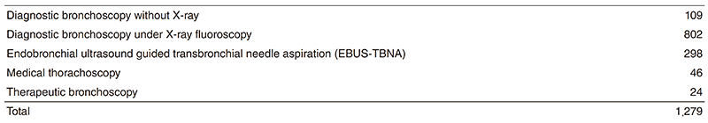
Research activities
Our efforts have been focused on new diagnostic and therapeutic strategies including bronchoscopy, which involve CT-screening for lung cancer and lead to cures and less-invasive treatments for lung cancer. To achieve a more accurate endoscopic diagnosis for solitary peripheral lung nodules, we are using threedimensional computed tomography (3DCT) navigation, ultrasound-guided approach, electromagnetic navigation and/or onsite cytology.
We showed the utility of a cryoprobe for biopsy during medical thoracoscopy. We also showed the utility of a bronchoscopic procedure for re-biopsy.
Education
A flexible bronchoscope was developed for the first time in the world in this institution. These are many residents and overseas doctors wishing to train at our division. We were given the opportunity of writing papers and conference presentations for many residents. Overseas training doctors came from many countries.
Future prospects
Development of a new diagnostic method with a compact electron microscope and a multicenter clinical trial for evaluation of photodynamic therapy for peripheral lung cancer are expected to be carried out. We are also planning a study for utility of radiomics as a tool for diagnosis and as a criterion for management of peripheral lung nodules.
List of papers published in 2018
Journal
1. Nakai T, Matsumoto Y, Sasada S, Tanaka M, Tsuchida T, Ohe Y, Motoi N. Cryobiopsy during flex-rigid pleuroscopy: an emerging alternative biopsy method in malignant pleural mesothelioma A comparative study of pathology. Jpn J Clin Oncol, 49:559-566, 2019
2. Ohashi-Nakatani K, Shibuki Y, Fujima M, Watanabe R, Yoshida A, Yoshida H, Matsumoto Y, Tsuchida T, Watanabe SI, Motoi N. Primary pulmonary meningioma: A rare case report of aspiration cytological features and immunohistochemical assessment. Diagn Cytopathol, 47:330-333, 2019
3. Hiraishi Y, Izumo T, Sasada S, Matsumoto Y, Nakai T, Tsuchida T, Baba H. Factors affecting bacterial culture positivity in specimens from bronchoscopy in patients with suspected lung cancer. Respir Investig, 56:457-463, 2018