Annual Report 2018
Department of Pathology and Clinical Laboratories
Nobuyoshi Hiraoka, Shigeki Sekine, Hiromichi Matsushita, Noriko Motoi, Akiko Maeshima, Taisuke Mori, Hirokazu Taniguchi, Reiko Watanabe, Masayuki Yoshida, Akihiko Yoshida, Hiroshi Yoshida, Kaishi Satomi, Kuniko Sunami, Taiki Hashimoto
Introduction
The practice, education, and research on diagnostic and anatomic pathology are carried out in the Pathology Division. Diagnostic pathology practice comprises all issues concerning the processing of cell and tissue specimens obtained from patients, preparation of tissue blocks and pathology slides, and histological and cytopathological diagnoses of diseases. Anatomic pathology practice, by contrast, comprises the autopsy and post-mortem systemic gross and microscopic examinations of patients. Case conferences with each clinical division are held periodically, and residents and trainees are accepted for training in diagnostic pathology on a rotating basis. To provide more accurate and informative diagnoses in the future, the staff members conduct basic, clinical, or translational research by themselves or in collaboration with other divisions or institutions.
The Clinical Laboratories Division provides an important service as an in-hospital diagnostic unit by examining laboratory specimens and screening for disorders. All laboratory data are provided for clinicians under strict internal and external quality control. The laboratories in this department have acquired ISO 15189 accreditation, which certifies the quality and competency of a medical laboratory with regard to the quality management and technique, as established by the International Organization for Standardization's Technical Committee 212 (ISO/ TC 212). The staff of the Clinical Laboratories Division is continuously striving to maintain the quality and quantity of laboratory services through this system.
The Team and What We Do
Pathology Division:
In 2018, 12 staff pathologists, seven residents, and 14 medical technologists, including 12 cytotechnologists, cooperatively performed routine histological and cytopathological diagnoses of specimens obtained from patients at the National Cancer Center Hospital (NCCH) and educated residents. We accepted a total of 25,502 histological specimens, including 20,806 biopsy and 4,696 surgically resected specimens and 12,731 cytological specimens (Table 1). These included 1,969 intraoperative frozen sections and 342 intraoperative cytological diagnoses. We also provided a total of 192 pathological diagnoses to the outpatient clinic for pathology consultation (second opinion).
Clinical Laboratories Division:
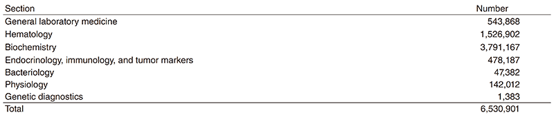
A total of 56 full-time and 13 part-time medical technologists, two photographers, and five assistants provide services in this division. These staff work in the sections of 1) general laboratory medicine and hematology, 2) biochemistry, 3) endocrinology, immunology, and tumor markers, 4) bacteriology, 5) genetic diagnostics, 6) transfusion, 7) phlebotomy, 8) physiological examination, and 9) pathology in the NCCH and in the sections of phlebotomy and physiological examination in the Screening Center. Sections 1) to 5) are supervised by Drs. Hiromichi Matsushita and Kuniko Sunami; 6) and 7) by Dr. Saiko Kurosawa (Transfusion Therapy); 8) by Dr. Yasunori Mizuguchi (Diagnostic Radiology), Drs. Masaaki Shoji and Takeshi Iwasa (General Internal Medicine), and Dr. Eriko Iwamoto (Breast Surgery), Drs. Fumihiko Matsumoto, Masahiko Fukasawa, Go Omura, Kenya Kobayashi, Satoko Matsumura and Atsuo Ikeda (Head and Neck Oncology); and 9) by the doctors in the Pathology Division. The bacteriology staff are members of the Infection Control Team and participate in infection management. The actual number of laboratory tests performed in this division in 2018 is shown in Table 2.
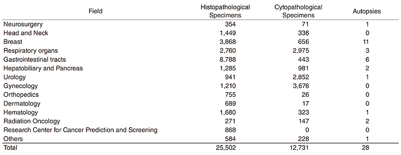
Table 2. Number of laboratory tests examined in the Clinical Laboratories Division in 2018
Research activities
1. Hepato-biliary pancreatic pathology
We reported that the intrapancreatic neural density is reduced in pancreatic cancer tissues and an independent prognosticator. The frequency of HER2 positivity was found to differ among the organs in the biliary tract, and the HER2 protein expression was usually heterogenous.
2. Gastrointestinal pathology
We demonstrated the frequent presence of RHOA mutations in intestinal-type adenocarcinoma with anastomosing glands of the stomach, suggesting this is a genetically distinct subtype. Our analysis of precursor polyp-associated traditional serrated adenomas (TSAs) revealed the role of the WNT pathway gene mutation in the development of TSA. We proposed superficially serrated adenoma as a novel subtype of colorectal serrated lesion. An analysis of a large number of sessile serrated adenoma/polyps revealed a degree of genetic heterogeneity of this entity.
3. Thoracic pathology
We conducted clinicopathological, morphological, and genetic analyses to identify predictive factors of a prognostic or therapeutic response in cases of lung and other thoracic malignancies. We reported the clinicopathological significance of a histopathological assessment of invasive mucinous adenocarcinoma (a rare variant of lung cancer) related to the outcome, a clinicopathological comparison study between lung and mediastinal neuroendocrine tumor, and a feasibility study of lung cytology specimens for biomarker tests. We also contributed to several collaboration studies, including a study of the SP gene mutation in idiopathic pulmonary fibrosisassociated non-small cell carcinoma and a study on the clinical use of cytology specimens for biomarker tests.
4. Bone and soft tissue pathology
We revisited the utility of MDM2 and H3K27me3 in differentiating malignant peripheral nerve sheath tumor and dedifferentiated liposarcoma. The MDM2 and H3K27me3 status was also examined in extraskeletal osteosarcoma to evaluate the disease heterogeneity. We showed that INSM1 is often expressed in extraskeletal myxoid chondrosarcoma, which may have potential diagnostic utility. We compared the utility of NKX2-2 and PAX7 in the diagnosis of Ewing sarcoma and discovered that a subset of dedifferentiated chondrosarcoma is deficient in H3K27me3, and this group is associated with characteristic clinicopathological features. We contributed to the comprehensive genetic and epigenetic analyses of myxofibrosarcoma and an international project to identify the fusioncreating mechanism in Ewing sarcoma.
5. Breast pathology
We reported our experience with two cases of endosalpingiosis in axillary SLNs, which we confirmed using immunohistochemistry. We also reported biomarker discordance between primary breast cancer and bone or bone marrow metastases.
6. Gynecological pathology
We reported positive pelvic cytology as a prognostic factor for stage I-II uterine serous carcinoma. We also showed that synchronous endometrial and ovarian endometrioid carcinoma (SEO-EC) patients had unique prognostic features, indicating that SEO-EC patients with tumors localized to the uterine body and adnexa lesions had a low risk for recurrence and might not require adjuvant therapy. We reported the prevalence of lymphangioleiomyomatosis in lymph nodes sampled in gynecological surgery and discussed the risk of subsequent pulmonary LAM. We reported a case of endometrial cancer among patients with Cornelia de Lange syndrome and a case of endometrial cancer showing a unique morphology and tumor extension in a patient with Cowden syndrome. We and the researchers of NCCRI have collaborated on several research projects and reported targets of synthetic lethality in ovarian clear cell carcinoma with an ARID1A mutation.
7. Head and neck/ophthalmic pathology
We reported that the CyclinD1 expression in thyroid nodules is a novel marker for both the sensitivity and specificity and that a screening system using CyclinD1 is also useful for cytopathology. We analyzed p53 protein expression and all TP53 exon sequences in 317 head and neck squamous cell carcinomas. We classified two risk categories as "p53 adverse function" and "p53 favorable function" based on TP53 mutation status and p53 protein phenotype. The "p53 adverse function" was an independent poor predictor of overall survival, local control, and distant metastasis-free survival.
8. Brain tumor pathology
We developed a novel FISH probe set to improve the diagnostic accuracy of 1p/19q codeleted glioma in collaboration with the Division of Brain Tumor Translational Research. The utility of H3K27me3 immunohistochemistry for dividing posterior fossa ependymomas into PFA and PFB groups was confirmed. We showed that IDH1 (R132H) mutation-specific antibody often provides false-negative results in frozen section control slides.
9. Skin pathology
We reported the frequent presence of transforming YAP1 fusions (YAP1-MAML2, YAP1-NUTM1, WWTR1-NUTM1) in poromas and porocarcinoma. The YAP1 and WWTR1 fusions strongly transactivated a TEAD reporter and promoted anchorage-independent growth, confirming their tumorigenic roles. These findings suggest that YAP1/TEAD-dependent transcription is a candidate therapeutic target for porocarcinoma.
10. Clinical laboratories
In order to facilitate the implementation of cancer genome medicine, wherein gene mutation profiles in cancer tissues are used to select ideal treatment options, we performed Advanced Medical Care B clinical research using the NCC Oncopanel System. Several medical technologists obtained interesting findings in their routine practice and made presentations at several domestic medical conferences. In the cytogenetics section, the FISH technique using the Metafer system to evaluate the gene amplification of HER2; the gene rearrangement of DDIT3, EWSR1 and SS18 for musculoskeletal/soft tissue tumors; and the formation of IGH-MYC and IGH-BCL2 for malignant lymphoma provided useful data for not only clinical practices but also research activities of doctors in the NCCH and NCCRI. In addition, an in-hospital bio-bank system has been maintained in order to aid in clinical research being conducted by various researchers in the NCCH and NCCRI. More than 600,000 postclinical-test serum samples have been stored at -20°C as of the end of March 2019.
List of papers published in 2018
Journal
1. Nakano Y, Tomiyama A, Kohno T, Yoshida A, Yamasaki K, Ozawa T, Fukuoka K, Fukushima H, Inoue T, Hara J, Sakamoto H, Ichimura K. Identification of a novel KLC1-ROS1 fusion in a case of pediatric low-grade localized glioma. Brain Tumor Pathol, 36:14-19, 2019
2. Asano N, Matsuzaki J, Ichikawa M, Kawauchi J, Takizawa S, Aoki Y, Sakamoto H, Yoshida A, Kobayashi E, Tanzawa Y, Nakayama R, Morioka H, Matsumoto M, Nakamura M, Kondo T, Kato K, Tsuchiya N, Kawai A, Ochiya T. A serum microRNA classifier for the diagnosis of sarcomas of various histological subtypes. Nat Commun, 10:1299, 2019
3. Hashimoto T, Ogawa R, Tang TY, Yoshida H, Taniguchi H, Katai H, Oda I, Sekine S. RHOA mutations and CLDN18-ARHGAP fusions in intestinal-type adenocarcinoma with anastomosing glands of the stomach. Mod Pathol, 32:568-575, 2019
4. Hashimoto T, Ogawa R, Yoshida H, Taniguchi H, Kojima M, Saito Y, Sekine S. Acquisition of WNT Pathway Gene Alterations Coincides With the Transition From Precursor Polyps to Traditional Serrated Adenomas. Am J Surg Pathol, 43:132-139, 2019
5. Hironaka-Mitsuhashi A, Tsuda H, Yoshida M, Shimizu C, Asaga S, Hojo T, Tamura K, Kinoshita T, Ushijima T, Hiraoka N, Fujiwara Y. Invasive breast cancers in adolescent and young adult women show more aggressive immunohistochemical and clinical features than those in women aged 40-44 years. Breast Cancer, 26:386-396, 2019
6. Ino Y, Oguro S, Yamazaki-Itoh R, Hori S, Shimada K, Hiraoka N. Reliable evaluation of tumor-infiltrating lymphocytes in pancreatic cancer tissue biopsies. Oncotarget, 10:1149-1159, 2019
7. Jain D, Nambirajan A, Borczuk A, Chen G, Minami Y, Moreira AL, Motoi N, Papotti M, Rekhtman N, Russell PA, Savic Prince S, Yatabe Y, Bubendorf L. Immunocytochemistry for predictive biomarker testing in lung cancer cytology. Cancer Cytopathol, 127:325-339, 2019
8. Kawai H, Matsushita H, Kawakami S, Furuya D, Shiraiwa-Hara S, Ichiki A, Hara R, Aoyama Y, Ogiya D, Suzuki R, Machida S, Onizuka M, Shirasugi Y, Ogawa Y, Kawada H, Nakamura N, Ando K. A Case of Composite Lymphoma with Extranodal NK/T-cell Lymphoma, Nasal-type and Diffuse Large B-cell Lymphoma. J Clin Exp Hematop, 59:34-39, 2019
9. Kobayashi K, Yoshimoto S, Matsumoto F, Ando M, Murakami N, Omura G, Fukasawa M, Matsumoto Y, Matsumura S, Akamatsu M, Hiraoka N, Eigitsu R, Mori T. All-Exon TP53 Sequencing and Protein Phenotype Analysis Accurately Predict Clinical Outcome after Surgical Treatment of Head and Neck Squamous Cell Carcinoma. Ann Surg Oncol, 26:2294-2303, 2019
10. Kojima N, Yoshida H, Uehara T, Ushigusa T, Asami Y, Shiraishi K, Kato T. Primary Clear Cell Adenocarcinoma of the Vulva: A Case Study With Mutation Analysis and Literature Review. Int J Surg Pathol, 1066896919848823, 2019.
11. Kuno I, Tate K, Yoshida H, Takahashi K, Kato T. Endometrial Endometrioid Carcinoma With Ovarian Metastasis Showing Morula-like Features in a Patient With Cowden Syndrome: A Case Report. Int J Gynecol Pathol, 2019
12. Kuno I, Yoshida H, Kohno T, Ochiai A, Kato T. Endometrial cancer arising after complete remission of uterine malignant lymphoma: A case report and mutation analysis. Gynecol Oncol Rep, 28:50- 53, 2019
13. Makise N, Sekimizu M, Konishi E, Motoi T, Kubo T, Ikoma H, Watanabe SI, Okuma T, Hiraoka N, Fukayama M, Kawai A, Ichikawa H, Yoshida A. H3K27me3 deficiency defines a subset of dedifferentiated chondrosarcomas with characteristic clinicopathological features. Mod Pathol, 32:435-445, 2019
14. Nakai T, Matsumoto Y, Sasada S, Tanaka M, Tsuchida T, Ohe Y, Motoi N. Cryobiopsy during flex-rigid pleuroscopy: an emerging alternative biopsy method in malignant pleural mesothelioma. A comparative study of pathology. Jpn J Clin Oncol, 49:559-566, 2019
15. Nakamura Y, Shida D, Shibayama T, Yoshida A, Matsui Y, Shinoda Y, Iwata S, Kanemitsu Y. Giant multilocular prostatic cystadenoma. World J Surg Oncol, 17:42, 2019
16. Ogasawara A, Matsushita H, Tanaka Y, Shirasugi Y, Ando K, Asai S, Miyachi H. A simple screening method for the diagnosis of chronic myeloid leukemia using the parameters of a complete blood count and differentials. Clin Chim Acta, 489:249-253, 2019
17. Ogiwara H, Takahashi K, Sasaki M, Kuroda T, Yoshida H, Watanabe R, Maruyama A, Makinoshima H, Chiwaki F, Sasaki H, Kato T, Okamoto A, Kohno T. Targeting the Vulnerability of Glutathione Metabolism in ARID1A-Deficient Cancers. Cancer Cell, 35:177-190.e8, 2019
18. Ohashi-Nakatani K, Shibuki Y, Fujima M, Watanabe R, Yoshida A, Yoshida H, Matsumoto Y, Tsuchida T, Watanabe SI, Motoi N. Primary pulmonary meningioma: A rare case report of aspiration cytological features and immunohistochemical assessment. Diagn Cytopathol, 47:330-333, 2019
19. Oyama R, Kito F, Qiao Z, Sakumoto M, Shiozawa K, Toki S, Yoshida A, Kawai A, Kondo T. Establishment of novel patient-derived models of dermatofibrosarcoma protuberans: two cell lines, NCC-DFSP1-C1 and NCC-DFSP2-C1. In Vitro Cell Dev Biol Anim, 55:62-73, 2019
20. Sato J, Shimoi T, Shimomura A, Noguchi E, Kodaira M, Yunokawa M, Yonemori K, Shimizu C, Fujiwara Y, Yoshida M, Tamura K. The Incidence of Nonmalignant Diseases among Patients with Suspected Carcinoma of Unknown Primary Site. Intern Med, 58:1423-1428, 2019
21. Sekimizu M, Yoshida A, Mitani S, Asano N, Hirata M, Kubo T, Yamazaki F, Sakamoto H, Kato M, Makise N, Mori T, Yamazaki N, Sekine S, Oda I, Watanabe SI, Hiraga H, Yonemoto T, Kawamoto T, Naka N, Funauchi Y, Nishida Y, Honoki K, Kawano H, Tsuchiya H, Kunisada T, Matsuda K, Inagaki K, Kawai A, Ichikawa H. Frequent mutations of genes encoding vacuolar H+ -ATPase components in granular cell tumors. Genes Chromosomes Cancer, 58:373-380, 2019
22. Shiino S, Yoshida M, Jimbo K, Asaga S, Takayama S, Maeshima A, Tsuda H, Kinoshita T, Hiraoka N. Two rare cases of endosalpingiosis in the axillary sentinel lymph nodes: evaluation of immunohistochemical staining and one-step nucleic acid amplification (OSNA) assay in patients with breast cancer. Virchows Arch, 474:633-638, 2019
23. Tate K, Yoshida H, Ishikawa M, Shimizu H, Uehara T, Kato T. Endometrial Carcinoma With an Unusual Morphology in a Patient With Cornelia de Lange Syndrome: A Case Study. Int J Gynecol Pathol, 38:340-345, 2019
24. Teshima M, Tokita K, Ryo E, Matsumoto F, Kondo M, Ikegami Y, Shinomiya H, Otsuki N, Hiraoka N, Nibu KI, Yoshimoto S, Mori T. Clinical impact of a cytological screening system using cyclin D1 immunostaining and genomic analysis for the diagnosis of thyroid nodules. BMC Cancer, 19:245, 2019
25. Tozawa K, Ono-Uruga Y, Yazawa M, Mori T, Murata M, Okamoto S, Ikeda Y, Matsubara Y. Megakaryocytes and platelets from a novel human adipose tissue-derived mesenchymal stem cell line. Blood, 133:633-643, 2019
26. Uehara T, Yoshida H, Tate K, Kato T. Metachronous occurrence of two different histological subtypes of endometriosis-related neoplasms. Gynecol Oncol Rep, 27:42-45, 2019
27. Yatabe Y, Dacic S, Borczuk AC, Warth A, Russell PA, Lantuejoul S, Beasley MB, Thunnissen E, Pelosi G, Rekhtman N, Bubendorf L, Mino-Kenudson M, Yoshida A, Geisinger KR, Noguchi M, Chirieac LR, Bolting J, Chung JH, Chou TY, Chen G, Poleri C, Lopez-Rios F, Papotti M, Sholl LM, Roden AC, Travis WD, Hirsch FR, Kerr KM, Tsao MS, Nicholson AG, Wistuba I, Moreira AL. Best Practices Recommendations for Diagnostic Immunohistochemistry in Lung Cancer. J Thorac Oncol, 14:377-407, 2019
28. Yoneoka Y, Yoshida H, Ishikawa M, Shimizu H, Uehara T, Murakami T, Kato T. Prognostic factors of synchronous endometrial and ovarian endometrioid carcinoma. J Gynecol Oncol, 30:e7, 2019
29. Yoshida A, Satomi K, Ohno M, Matsushita Y, Takahashi M, Miyakita Y, Hiraoka N, Narita Y, Ichimura K. Frequent false-negative immunohistochemical staining with IDH1 (R132H)-specific H09 antibody on frozen section control slides: a potential pitfall in glioma diagnosis. Histopathology, 74:350-354, 2019
30. Yuda S, Shimizu C, Yoshida M, Shiino S, Kinoshita T, Maeshima AM, Tamura K. Biomarker discordance between primary breast cancer and bone or bone marrow metastases. Jpn J Clin Oncol, 49:426-430, 2019
31. Sunami K, Ichikawa H, Kubo T, Kato M, Fujiwara Y, Shimomura A, Koyama T, Kakishima H, Kitami M, Matsushita H, Furukawa E, Narushima D, Nagai M, Taniguchi H, Motoi N, Sekine S, Maeshima A, Mori T, Watanabe R, Yoshida M, Yoshida A, Yoshida H, Satomi K, Sukeda A, Hashimoto T, Shimizu T, Iwasa S, Yonemori K, Kato K, Morizane C, Ogawa C, Tanabe N, Sugano K, Hiraoka N, Tamura K, Yoshida T, Fujiwara Y, Ochiai A, Yamamoto N, Kohno T. Feasibility and utility of a panel testing for 114 cancer-associated genes in a clinical setting: A hospital-based study. Cancer Sci, 110:1480-1490, 2019
32. Shiino S, Matsuzaki J, Shimomura A, Kawauchi J, Takizawa S, Sakamoto H, Aoki Y, Yoshida M, Tamura K, Kato K, Kinoshita T, Kitagawa Y, Ochiya T. Serum miRNA-based Prediction of Axillary Lymph Node Metastasis in Breast Cancer. Clin Cancer Res, 25:1817-1827, 2019
33. Kato M, Nakamura H, Nagai M, Kubo T, Elzawahry A, Totoki Y, Tanabe Y, Furukawa E, Miyamoto J, Sakamoto H, Matsumoto S, Sunami K, Arai Y, Suzuki Y, Yoshida T, Tsuchihara K, Tamura K, Yamamoto N, Ichikawa H, Kohno T, Shibata T. A computational tool to detect DNA alterations tailored to formalin-fixed paraffin-embedded samples in cancer clinical sequencing. Genome Med, 10:44, 2018
34. Ohara K, Arai E, Takahashi Y, Fukamachi Y, Ito N, Maeshima AM, Fujimoto H, Yoshida T, Kanai Y. Feasibility of methylome analysis using small amounts of genomic DNA from formalin-fixed paraffin-embedded tissue. Pathol Int, 68:633-635, 2018
35. Anderson ND, de Borja R, Young MD, Fuligni F, Rosic A, Roberts ND, Hajjar S, Layeghifard M, Novokmet A, Kowalski PE, Anaka M, Davidson S, Zarrei M, Id Said B, Schreiner LC, Marchand R, Sitter J, Gokgoz N, Brunga L, Graham GT, Fullam A, Pillay N, Toretsky JA, Yoshida A, Shibata T, Metzler M, Somers GR, Scherer SW, Flanagan AM, Campbell PJ, Schiffman JD, Shago M, Alexandrov LB, Wunder JS, Andrulis IL, Malkin D, Behjati S, Shlien A. Rearrangement bursts generate canonical gene fusions in bone and soft tissue tumors. Science, 361:2018
36. Ando Y, Maeshima AM, Fukuhara S, Makita S, Munakata W, Suzuki T, Maruyama D, Taniguchi H, Izutsu K. CD3+ CD56+ EBER1+ atypical extraosseous plasmacytoma of the nasal cavity. Int J Hematol, 108:344-347, 2018
37. Cho H, Hashimoto T, Yoshida H, Taniguchi H, Ogawa R, Mori T, Hiraoka N, Saito Y, Sekine S. Reappraisal of the genetic heterogeneity of sessile serrated adenoma/polyp. Histopathology, 73:672-680, 2018
38. Ebata T, Shimoi T, Bun S, Miyake M, Yoshida A, Shimomura A, Noguchi E, Yonemori K, Shimizu C, Fujiwara Y, Narita Y, Tamura K. Efficacy and Safety of Pazopanib for Recurrent or Metastatic Solitary Fibrous Tumor. Oncology, 94:340-344, 2018
39. Frezza AM, Jones RL, Lo Vullo S, Asano N, Lucibello F, Ben-Ami E, Ratan R, Teterycz P, Boye K, Brahmi M, Palmerini E, Fedenko A, Vincenzi B, Brunello A, Desar IME, Benjamin RS, Blay JY, Broto JM, Casali PG, Gelderblom H, Grignani G, Gronchi A, Hall KS, Mir O, Rutkowski P, Wagner AJ, Anurova O, Collini P, Dei Tos AP, Flucke U, Hornick JL, Lobmaier I, Philippe T, Picci P, Ranchere D, Renne SL, Sbaraglia M, Thway K, Wagrodzki M, Wang WL, Yoshida A, Mariani L, Kawai A, Stacchiotti S. Anthracycline, Gemcitabine, and Pazopanib in Epithelioid Sarcoma: A Multi-institutional Case Series. JAMA Oncol, 4:e180219, 2018
40. Hama N, Totoki Y, Miura F, Tatsuno K, Saito-Adachi M, Nakamura H, Arai Y, Hosoda F, Urushidate T, Ohashi S, Mukai W, Hiraoka N, Aburatani H, Ito T, Shibata T. Epigenetic landscape influences the liver cancer genome architecture. Nat Commun, 9:1643, 2018
41. Ito Y, Makita S, Maeshima AM, Hatta S, Suzuki T, Yuda S, Fukuhara S, Munakata W, Suzuki T, Maruyama D, Izutsu K. Paraneoplastic Pemphigus Associated with B-cell Chronic Lymphocytic Leukemia Treated with Ibrutinib and Rituximab. Intern Med, 57:2395-2398, 2018
42. Karakawa A, Taoka K, Kaburaki T, Tanaka R, Shinozaki-Ushiku A, Hayashi H, Miyagi-Maeshima A, Nishimura Y, Uekusa T, Kojima Y, Fukayama M, Kurokawa M, Aihara M. Clinical features and outcomes of secondary intraocular lymphoma. Br J Haematol, 183:668-671, 2018
43. Kawai H, Matsushita H, Aoyama Y, Matsui K, Onizuka M, Ando K. Dysplastic features seen in a patient with acute myeloid leukemia harboring the KMT2A-TET1 fusion gene. Int J Hematol, 108:1-2, 2018
44. Kishi Y, Nara S, Esaki M, Hiraoka N, Shimada K. Extent of lymph node dissection in patients with gallbladder cancer. Br J Surg, 105:1658-1664, 2018
45. Kito F, Oyama R, Sakumoto M, Takahashi M, Shiozawa K, Qiao Z, Sakamoto H, Hirose T, Setsu N, Yoshida A, Kawai A, Kondo T. Establishment and characterization of novel patient-derived osteosarcoma xenograft and cell line. In Vitro Cell Dev Biol Anim, 54:528-536, 2018
46. Kuno I, Yoshida H, Shimizu H, Uehara T, Uno M, Ishikawa M, Kato T. Incidental lymphangioleiomyomatosis in the lymph nodes of gynecologic surgical specimens. Eur J Obstet Gynecol Reprod Biol, 231:93-97, 2018
47. Machado I, Yoshida A, Morales MGN, Abrahao-Machado LF, Navarro S, Cruz J, Lavernia J, Parafioriti A, Picci P, Llombart-Bosch A. Review with novel markers facilitates precise categorization of 41 cases of diagnostically challenging, "undifferentiated small round cell tumors". A clinicopathologic, immunophenotypic and molecular analysis. Ann Diagn Pathol, 34:1-12, 2018
48. Makise N, Sekimizu M, Kubo T, Wakai S, Watanabe SI, Kato T, Kinoshita T, Hiraoka N, Fukayama M, Kawai A, Ichikawa H, Yoshida A. Extraskeletal osteosarcoma: MDM2 and H3K27me3 analysis of 19 cases suggest disease heterogeneity. Histopathology, 73:147-156, 2018
49. Makita S, Maeshima AM, Maruyama D, Izutsu K, Tobinai K. Forodesine in the treatment of relapsed/refractory peripheral T-cell lymphoma: an evidence-based review. Onco Targets Ther, 11:2287-2293, 2018
50. Masai K, Motoi N. Response to Letter to the Editor Titled "Comments on Prognostic Impact of Margin Distance and Tumor Spread through Air Spaces in Limited Resection for Primary Lung Cancer". J Thorac Oncol, 13:e58-e59, 2018
51. Matsuo K, Takazawa Y, Ross MS, Elishaev E, Yunokawa M, Sheridan TB, Bush SH, Klobocista MM, Blake EA, Takano T, Baba T, Satoh S, Shida M, Ikeda Y, Adachi S, Yokoyama T, Takekuma M, Yanai S, Takeuchi S, Nishimura M, Iwasaki K, Johnson MS, Yoshida M, Hakam A, Machida H, Mhawech-Fauceglia P, Ueda Y, Yoshino K, Kajiwara H, Hasegawa K, Yasuda M, Miyake TM, Moriya T, Yuba Y, Morgan T, Fukagawa T, Pejovic T, Nagano T, Sasaki T, Richmond AM, Post MD, Shahzad MMK, Im DD, Yoshida H, Enomoto T, Omatsu K, Ueland FR, Kelley JL, Karabakhtsian RG, Roman LD. Proposal for a Risk-Based Categorization of Uterine Carcinosarcoma. Ann Surg Oncol, 25:3676-3684, 2018
52. Matsuo K, Takazawa Y, Ross MS, Elishaev E, Yunokawa M, Sheridan TB, Bush SH, Klobocista MM, Blake EA, Takano T, Baba T, Satoh S, Shida M, Ikeda Y, Adachi S, Yokoyama T, Takekuma M, Yanai S, Takeuchi S, Nishimura M, Iwasaki K, Johnson MS, Yoshida M, Hakam A, Machida H, Mhawech-Fauceglia P, Ueda Y, Yoshino K, Kajiwara H, Hasegawa K, Yasuda M, Miyake TM, Moriya T, Yuba Y, Morgan T, Fukagawa T, Pejovic T, Nagano T, Sasaki T, Richmond AM, Post MD, Shahzad MMK, Im DD, Yoshida H, Enomoto T, Omatsu K, Ueland FR, Kelley JL, Karabakhtsian RG, Roman LD. Significance of Lymphovascular Space Invasion by the Sarcomatous Component in Uterine Carcinosarcoma. Ann Surg Oncol, 25:2756-2766, 2018
53. Miura A, Mori T, Yoshida A, Watanabe Y, Sunami K, Watanabe S, Kohno T, Tsuta K. Primary adenocarcinoma of the trachea revealing a mucinous bronchial gland cell origin. Pathol Res Pract, 214:796-799, 2018
54. Nagumo Y, Maejima A, Toyoshima Y, Komiyama M, Yonemori K, Yoshida A, Fujimoto H. Neoadjuvant crizotinib in ALK-rearranged inflammatory myofibroblastic tumor of the urinary bladder: A case report. Int J Surg Case Rep, 48:1-4, 2018
55. Nomura T, Maki D, Matsumoto F, Mori T, Yoshimoto S. A rare case of coexisting lacrimal sac adenocarcinoma and transitional cell carcinoma. Ear Nose Throat J, 97:E32-E35, 2018
56. Ogura K, Hosoda F, Arai Y, Nakamura H, Hama N, Totoki Y, Yoshida A, Nagai M, Kato M, Arakawa E, Mukai W, Rokutan H, Kawai A, Tanaka S, Shibata T. Integrated genetic and epigenetic analysis of myxofibrosarcoma. Nat Commun, 9:2765, 2018
57. Oyama R, Kito F, Qiao Z, Sakumoto M, Noguchi R, Takahashi M, Toki S, Tanzawa Y, Yoshida A, Kawai A, Kondo T. Establishment of a novel patient-derived Ewing's sarcoma cell line, NCCES1-C1. In Vitro Cell Dev Biol Anim, 54:770-778, 2018
58. Oyama R, Kito F, Sakumoto M, Shiozawa K, Toki S, Endo M, Yoshida A, Kawai A, Kondo T. Establishment and proteomic characterization of a novel synovial sarcoma cell line, NCC-SS2-C1. In Vitro Cell Dev Biol Anim, 54:392-399, 2018
59. Oyama R, Takahashi M, Kito F, Sakumoto M, Shiozawa K, Qiao Z, Yoshida A, Endo M, Kawai A, Kondo T. Establishment and characterization of patient-derived xenograft and its cell line of primary leiomyosarcoma of bone. In Vitro Cell Dev Biol Anim, 54:458- 467, 2018
60. Sawada T, Hilhorst R, Rangarajan S, Yoshida M, Tanabe Y, Tamura K, Kinoshita T, Shimoyama T, van Beuningen R, Ruijtenbeek R, Tsuda H, Koizumi F. Inactive immune pathways in triple negative breast cancers that showed resistance to neoadjuvant chemotherapy as inferred from kinase activity profiles. Oncotarget, 9:34229-34239, 2018
61. Sekine K, Kanda S, Goto Y, Horinouchi H, Fujiwara Y, Yamamoto N, Motoi N, Ohe Y. Change in the lymphocyte-to-monocyte ratio is an early surrogate marker of the efficacy of nivolumab monotherapy in advanced non-small-cell lung cancer. Lung Cancer, 124:179-188, 2018
62. Shiota M, Naya M, Yamamoto T, Hishiki T, Tani T, Takahashi H, Kubo A, Koike D, Itoh M, Ohmura M, Kabe Y, Sugiura Y, Hiraoka N, Morikawa T, Takubo K, Suina K, Nagashima H, Sampetrean O, Nagano O, Saya H, Yamazoe S, Watanabe H, Suematsu M. Gold-nanofeve surface-enhanced Raman spectroscopy visualizes hypotaurine as a robust anti-oxidant consumed in cancer survival. Nat Commun, 9:1561, 2018
63. Sugiura Y, Kanda H, Motoi N, Nomura K, Inamura K, Okada E, Matsumoto H, Shimoji T, Matsumoto S, Nakayama J, Takazawa Y, Ishikawa Y, Machinami R. Osteosarcoma arising in fibrous dysplasia, confirmed by mutational analysis of GNAS gene. Pathol Res Pract, 214:318-324, 2018
64. Sunami K, Takahashi H, Tsuchihara K, Takeda M, Suzuki T, Naito Y, Sakai K, Dosaka-Akita H, Ishioka C, Kodera Y, Muto M, Wakai T, Yamazaki K, Yasui W, Bando H, Fujimoto Y, Fukuoka S, Harano K, Kawazoe A, Kimura G, Koganemaru S, Kogawa T, Kotani D, Kuboki Y, Matsumoto H, Matsumoto S, Mishima S, Nakamura Y, Sawada K, Shingaki S, Shitara K, Umemoto K, Umemura S, Yasuda K, Yoshino T, Yamamoto N, Nishio K. Clinical practice guidance for next-generation sequencing in cancer diagnosis and treatment (Edition 1.0). Cancer Sci, 109:2980-2985, 2018
65. Suzuki S, Sakurai H, Yotsukura M, Masai K, Asakura K, Nakagawa K, Motoi N, Watanabe SI. Clinical Features of Ground Glass Opacity-Dominant Lung Cancer Exceeding 3.0 cm in the Whole Tumor Size. Ann Thorac Surg, 105:1499-1506, 2018
66. Tsuchiya K, Tabe Y, Ai T, Ohkawa T, Usui K, Yuri M, Misawa S, Morishita S, Takaku T, Kakimoto A, Yang H, Matsushita H, Hanami T, Yamanaka Y, Okuzawa A, Horii T, Hayashizaki Y, Ohsaka A. Eprobe mediated RT-qPCR for the detection of leukemia-associated fusion genes. PLoS One, 13:e0202429, 2018
67. Tanaka Y, Maeshima AM, Nomoto J, Makita S, Fukuhara S, Munakata W, Maruyama D, Tobinai K, Kobayashi Y. Expression pattern of PD-L1 and PD-L2 in classical Hodgkin lymphoma, primary mediastinal large B-cell lymphoma, and gray zone lymphoma. Eur J Haematol, 100:511-517, 20187
68. Tate K, Watanabe R, Yoshida H, Shimizu H, Uehara T, Ishikawa M, Ikeda SI, Hiraoka N, Kato T. Uterine adenosarcoma in Japan: Clinicopathologic features, diagnosis and management. Asia Pac J Clin Oncol, 14:318-325, 2018
69. Tate K, Yoshida H, Ishikawa M, Uehara T, Ikeda SI, Hiraoka N, Kato T. Prognostic factors for patients with early-stage uterine serous carcinoma without adjuvant therapy. J Gynecol Oncol, 29:e34, 2018
70. Toki S, Motoi T, Miyake M, Kobayashi E, Kawai A, Yoshida A. Minute mesenchymal chondrosarcoma within osteochondroma: an unexpected diagnosis confirmed by HEY1-NCOA2 fusion. Hum Pathol, 81:255-260, 2018
71. Toki S, Wakai S, Sekimizu M, Mori T, Ichikawa H, Kawai A, Yoshida A. PAX7 immunohistochemical evaluation of Ewing sarcoma and other small round cell tumours. Histopathology, 73:645-652, 2018
72. Watanabe J, Makita S, Ito Y, Hatta S, Suzuki T, Yuda S, Maeshima AM, Fukuhara S, Munakata W, Suzuki T, Maruyama D, Kim SW, Izutsu K. Successful full-dose DeVIC therapy in a patient with advanced-stage extranodal natural killer/T-cell lymphoma refractory to L-asparaginase. Ann Hematol, 97:1739-1740, 2018
73. Yoshimura S, Munakata W, Ikeda C, Matsushita H. Marked erythroblastosis in myelodysplastic syndrome induced by gastric hemorrhaging. Int J Hematol, 107:387-389, 2018
74. Kito M, Munakata W, Ono K, Maeshima AM, Matsushita H. The infiltration of classical Hodgkin lymphoma cells into pleural effusion. Int J Hematol, 107:1-2, 2018
75. Yoshida H, Ikeda S, Tsukada T, Watanabe R, Hiraoka N, Kato T. Methods for Measuring and Staging a Uterine Cervical Adenocarcinoma Showing Intracystic Papillary Growth: A Case Report. Int J Gynecol Pathol, 37:364-367, 2018
76. Yoshida K, Fujiwara Y, Goto Y, Kohno T, Yoshida A, Tsuta K, Ohe Y. The first case of SMARCB1 (INI1) - deficient squamous cell carcinoma of the pleura: a case report. BMC Cancer, 18:398, 2018
77. Yoshimoto T, Motoi N, Yamamoto N, Nagano H, Ushijima M, Matsuura M, Okumura S, Yamaguchi T, Fukayama M, Ishikawa Y. Pulmonary Carcinoids and Low-Grade Gastrointestinal Neuroendocrine Tumors Show Common MicroRNA Expression Profiles, Different from Adenocarcinomas and Small Cell Carcinomas. Neuroendocrinology, 106:47-57, 2018
78. Aruga Y, Arakawa A, Ono K, Ogawa C, Matsushita H. Pseudo-Chediak-Higashi granules and Auer rods in mixed phenotype acute leukaemia, T/myeloid, not otherwise specified. Br J Haematol, 180:175, 2018
79. Nishimura Y, Yoshida A, Yonemori K, Motoi N, Tamura K, Hiraoka N, Mori T. SMARCB-1 deficient squamous cell carcinoma of a mediastinal cyst. Pathol Int, 68:563-566, 2018
80. Hashimoto T, Tanaka Y, Ogawa R, Mori T, Yoshida H, Taniguchi H, Hiraoka N, Kojima M, Oono Y, Saito Y, Sekine S. Superficially serrated adenoma: a proposal for a novel subtype of colorectal serrated lesion. Mod Pathol, 31:1588-1598, 2018
81. Shimoi T, Hamada A, Yamagishi M, Hirai M, Yoshida M, Nishikawa T, Sudo K, Shimomura A, Noguchi E, Yunokawa M, Yonemori K, Shimizu C, Kinoshita T, Fukuda T, Fujiwara Y, Tamura K. PIK3CA mutation profiling in patients with breast cancer, using a highly sensitive detection system. Cancer Sci, 109:2558-2566, 2018
82. Shimoi T, Yoshida M, Kitamura Y, Yoshino T, Kawachi A, Shimomura A, Noguchi E, Yunokawa M, Yonemori K, Shimizu C, Kinoshita T, Ichimura K, Fukuda T, Fujiwara Y, Tamura K. TERT promoter hotspot mutations in breast cancer. Breast Cancer, 25:292-296, 2018
83. Yamauchi T, Ohno M, Matsushita Y, Takahashi M, Miyakita Y, Kitagawa Y, Kondo E, Tsushita N, Satomi K, Yoshida A, Ichimura K, Narita Y. Radiological characteristics based on isocitrate dehydrogenase mutations and 1p/19q codeletion in grade II and III gliomas. Brain Tumor Pathol, 35:148-158, 2018
84. Miki S, Imamichi S, Fujimori H, Tomiyama A, Fujimoto K, Satomi K, Matsushita Y, Matsuzaki S, Takahashi M, Ishikawa E, Yamamoto T, Matsumura A, Mukasa A, Nishikawa R, Masutomi K, Narita Y, Masutani M, Ichimura K. Concomitant administration of radiation with eribulin improves the survival of mice harboring intracerebral glioblastoma. Cancer Sci, 109:2275-2285, 2018
85. Kobayashi K, Matsumoto F, Miyakita Y, Mori T, Shimoi T, Murakami N, Yoshida A, Arakawa A, Omura G, Fukasawa M, Matsumoto Y, Matsumura S, Itami J, Narita Y, Yoshimoto S. Impact of Surgical Margin in Skull Base Surgery for Head and Neck Sarcomas. J Neurol Surg B Skull Base, 79:437-444, 2018
86. Fukuoka K, Kanemura Y, Shofuda T, Fukushima S, Yamashita S, Narushima D, Kato M, Honda-Kitahara M, Ichikawa H, Kohno T, Sasaki A, Hirato J, Hirose T, Komori T, Satomi K, Yoshida A, Yamasaki K, Nakano Y, Takada A, Nakamura T, Takami H, Matsushita Y, Suzuki T, Nakamura H, Makino K, Sonoda Y, Saito R, Tominaga T, Matsusaka Y, Kobayashi K, Nagane M, Furuta T, Nakada M, Narita Y, Hirose Y, Ohba S, Wada A, Shimizu K, Kurozumi K, Date I, Fukai J, Miyairi Y, Kagawa N, Kawamura A, Yoshida M, Nishida N, Wataya T, Yamaoka M, Tsuyuguchi N, Uda T, Takahashi M, Nakano Y, Akai T, Izumoto S, Nonaka M, Yoshifuji K, Kodama Y, Mano M, Ozawa T, Ramaswamy V, Taylor MD, Ushijima T, Shibui S, Yamasaki M, Arai H, Sakamoto H, Nishikawa R, Ichimura K. Significance of molecular classification of ependymomas: C11orf95-RELA fusion-negative supratentorial ependymomas are a heterogeneous group of tumors. Acta Neuropathol Commun, 6:134, 2018