Annual Report 2018
Laboratory of Molecular Carcinogenesis
Naoto Tsuchiya, Yuko Nishiyama, Yuki Yoshimatsu, Takahiro Shirai, Yuma Nozue
Introduction
To develop new strategies for cancer therapy and diagnosis, understanding molecular networks, formed intercellularly and intracellularly, is a major theme to be tackled. Our laboratory has been focusing on the biological functions of non-coding RNAs (ncRNAs). microRNAs, a class of ncRNAs, are known to be strong regulators of intra- and intercellular connections of molecular networks. We are extensively analyzing tumor-related miRNAs to find intracellular networks required for carcinogenesis, and have a major interest in the extracellular miRNAs as regulators for cancer cells to adapt to their microenvironment.
Research activities
1) Identification of cellular tumor-suppressor network by using miRNAs
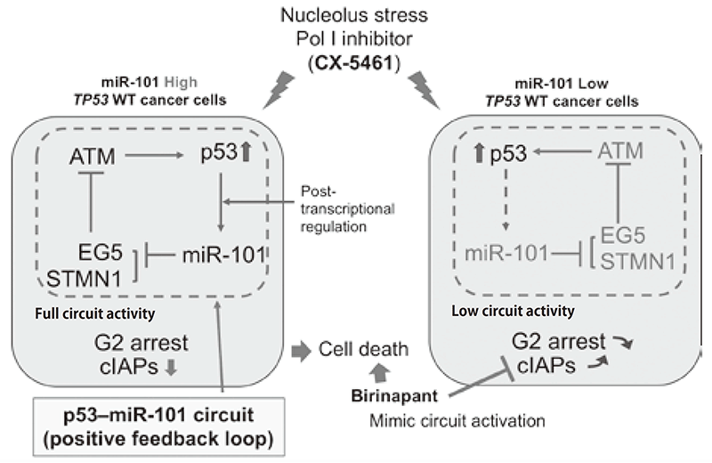
miR-101 was identified as a candidate tumorsuppressive miRNA. We discovered a molecular link between p53 and miR-101, which form a molecular circuit in response to nucleolar stress. miR-101 was post-transcriptionally activated after cells were exposed to RNA polymerase I inhibitor in a p53-dependent manner. miR-101 induces G2 phase-specific feedback regulation of p53 through the repression of EG5, resulting in the enhancement of apoptosis induction via activation of ATM-p53 signaling. In lung cancer patients, low expression of miR-101 was significantly associated with poor prognosis exclusively in p53 WT cases. Furthermore, the most downstream target of this circuit included the inhibitor of apoptosis proteins (IAPs). Repression of cIAPs by a selective inhibitor promoted apoptosis was induced by Pol I inhibitor in p53 WT cancer cells. Our results indicate that the molecular circuit formed by p53 and miR-101 is a crucial component of the cellular tumor-suppressor network induced by nucleolus stress and mimicking activation of p53-miR-101 circuit represents a promising strategy for cancer therapy (Figure 1, Fujiwara et al. EBioMedicine, 2018).
Figure 1. Regulation of p53-dependent nucleolus stress by miR-101
2) Regulation of Tumor-microenvironment (TME) by extracellular miRNA
Adaptation of sarcoma cells to their specific tumor microenvironment (TME) is a crucial component of maintenance of their malignant properties. Intracellular communication is generally mediated by cell-cell contact or secreted proteins, including cytokines and chemokine. Accumulating evidence demonstrates that extracellular vesicles (EVs), including those in exosomes and other particles, released by tumor cells play a major role in adaptation of malignant cells to TME. Extracellular microRNAs (exmiRNAs) embedded in the EVs have been reported to be involved in this adaptation, and also in the initiation of metastasis. We identified miR-451a as a candidate regulator of metastasis of sarcomas. miR-451a, whose high expression is associated with good prognosis of sarcoma patients, was identified by comprehensive expression analysis using clinical samples of sarcomas. Although miR-451a did not affect cell proliferation, it strongly repressed sarcoma cell migration. CMTM6 was identified as a target gene of miR-451a, and its repression induced changes in the expression levels of plasma membrane proteins, those included growth factor receptors. Furthermore, CMTM6 was found to localize to membrane structure of cells and secreted to the extracellular space. Interestingly, we found that CMTM6 vesicles contain a subset of miRNAs. Therefore, our data suggest that CMTM6- contining EVs is involved in the secretion of a subset of miRNAs and this function is a potent feature promoting maintenance of the malignant phenotypes of tumor cells.
3) Understanding the cellular networks specifically formed in p53-inactivated cancer cells
Tumor-suppressor gene, TP53, is mutated and/or inactivated in half of human cancers, and dysfunction of p53 protein, a gene product of TP53, makes a critical contribution to the onset of carcinogenesis. Molecular network(s) specifically activated in p53-deficient contexts may promote proliferation and survival of cancer cells. Therefore, understanding molecular bases for the activation of these networks provides the weaknesses of p53-deficient cancer cells, leading to the development of an ideal strategy for cancer therapy. We previously identified NEK9 as a regulator of the cell cycle in p53 deficient cells. NEK9 depression inhibited cell proliferation only in p53 deficient cells in vitro and in vivo. Parallel analyses identified a cellular network activated in p53 mutant context. Lung adenocarcinoma cases with mutation p53 showed significant upregulation of this cellular network, suggesting this network-dependent proliferation in p53 mutant cancer cells. Detailed molecular cross talks of NEK9 and this cellular network have been extensively analyzed.
Education
Supervising research and presentation skills for graduate school students and young scientists.
Future prospects
Our studies aim to clarify cellular network(s) formed in cancer cells. Results obtained from these studies will provide the molecular insights into how cancer cells were developed and progressed. Especially, research focusing on extracellular miRNA and extracellular vesicles is expected to find a novel molecular mechanism for intercellular communication between cancer cells and surrounding non-tumor cells, and this finding will be the new principle for the development of next cancer therapeutics.