Annual Report 2019
Division of Science and Technology for Endoscopy (Kashiwa Campus)
Hiroaki Ikematsu, Tomonori Yano, Toshihiro Takamatsu, Keisuke Hori, Takako Mamada, Eri Oishibashi
Introduction
The progress of endoscopic diagnosis and treatment equipment has been remarkable. However, further devices are expected to be developed in the future, such as cancer screening equipment, diagnostic equipment for cancers that are often overlooked, diagnostic equipment for deep lesions, and devices for treatment complications. Therefore, we are conducting basic research by incorporating the seeds of science and engineering academia into the development of these devices.
Research activities
Present research activities mainly focus on the development of new endoscopic imaging modalities. One project is the development of a diagnostic AI system for the detection of gastrointestinal lesions with the high efficiency of deep learning. Within this period, we succeeded in developing the AI model for the detection of stomach and colorectal lesions with a high accuracy rate (Figure 1). Another project is a new bioimaging system using near-infrared light with a wavelength of over 1,000 nm with various spectrums (Figure 1). This system is capable of penetrating through the gastrointestinal wall and obtaining images. Another project is examination of gastrointestinal lesion images in photoacoustic imaging (Figure 2). It may be possible to visualize deep vascular networks in the future. Other projects are development of an automatic colonoscope, development of a hemostatic device for endoscopes, and molecular biological analysis of early gastrointestinal cancer that underlies non-invasive diagnostics.
Figure 1. Identification of GIST using near-infrared hyperspectral imaging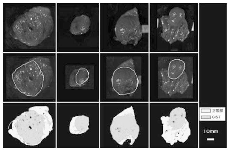
Figure 2. Deep gastrointestinal vascular images obtained using photoacoustic imaging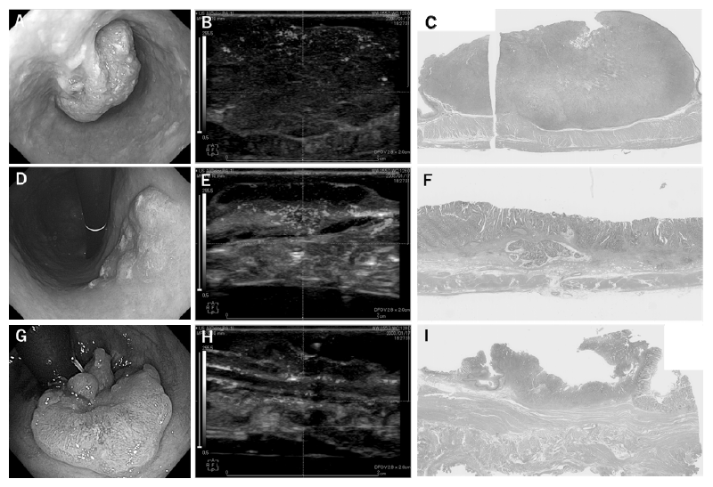
Clinical trials
1) Development of a diagnostic AI system for the detection of gastrointestinal lesions
2) Development of diagnostic endoscopy for GIST using near-infrared light
3) Photoacoustic imaging of fresh human surgical and endoscopic gastrointestinal specimens: a pilot study
Education
One researcher at Tokyo University of Science was sent to our hospital (National Cancer Center Hospital East: NCCHE) by a cross-appointment with Tokyo University of Science. Meetings are constantly conducted with faculties including technology and science students of Tokyo University of Science.
Future prospects
In the future, we will continue to develop endoscopic equipment in cooperation with the Department of Gastroenterology and Endoscopy of the NCCHE and try innovative approaches to produce all-new endoscopy in collaboration with academia and companies. With the aim of acquiring new seeds, we are developing the research infrastructure necessary to collaborate with academia.
List of papers published in 2019
Journal
1. Shinmura K, Ikematsu H, Kojima M, Nakamura H, Osera S, Yoda Y, Hori K, Oono Y, Ochiai A, Yano T. Safety of endoscopic procedures with monopolar versus bipolar instruments in an ex vivo porcine model. BMC Gastroenterol, 20:27, 2020
2. Takashima K, Fujii S, Komatsuzaki R, Komatsu M, Takahashi M, Kojima T, Daiko H, Minashi K, Chiwaki F, Muto M, Sasaki H, Yano T. CD24 and CK4 are upregulated by SIM2, and are predictive biomarkers for chemoradiotherapy and surgery in esophageal cancer. Int J Oncol, 56:835-847, 2020
3. Kadota T, Abe S, Yoda Y, Yoshinaga S, Oda I, Kojima T, Kato K, Daiko H, Yano T. Clinical outcomes according to the modified endoscopic criteria for neoadjuvant chemotherapy in resectable esophageal squamous cell carcinoma. Dig Endosc, 32:337-345, 2020
4. Minamide T, Yoda Y, Hori K, Shinmura K, Oono Y, Ikematsu H, Yano T. Advantages of salvage photodynamic therapy using talaporfin sodium for local failure after chemoradiotherapy or radiotherapy for esophageal cancer. Surg Endosc, 34:899-906, 2020
5. Yano T, Hasuike N, Ono H, Boku N, Ogawa G, Kadota T, Oda I, Doyama H, Hori S, Iishi H, Takahashi A, Takizawa K, Muto M. Factors associated with technical difficulty of endoscopic submucosal dissection for early gastric cancer that met the expanded indication criteria: post hoc analysis of a multi-institutional prospective confirmatory trial (JCOG0607). Gastric Cancer, 23:168-174, 2020
6. Yano T, Wang KK. Photodynamic Therapy for Gastrointestinal Cancer. Photochem Photobiol, 96:517-523, 2020
7. Sato D, Motegi A, Kadota T, Kojima T, Bando H, Shinmura K, Hori K, Yoda Y, Oono Y, Zenda S, Ikematsu H, Akimoto T, Yano T. Therapeutic results of proton beam therapy with concurrent chemotherapy for cT1 esophageal cancer and salvage endoscopic therapy for local recurrence. Esophagus, 17:305-311, 2020
8. Yamamoto Y, Kadota T, Yoda Y, Hori K, Hatogai K, Kojima T, Fujii S, Akimoto T, Yano T. Review of early endoscopic findings in patients with local recurrence after definitive chemoradiotherapy for esophageal squamous cell carcinoma. Esophagus, 2020
9. Kadota T, Yoda Y, Hori K, Shinmura K, Oono Y, Ikematsu H, Yano T. Prophylactic steroid administration against strictures is not enough for mucosal defects involving the entire circumference of the esophageal lumen after esophageal endoscopic submucosal dissection (ESD). Esophagus, 2020
10. Kudo SE, Misawa M, Mori Y, Hotta K, Ohtsuka K, Ikematsu H, Saito Y, Takeda K, Nakamura H, Ichimasa K, Ishigaki T, Toyoshima N, Kudo T, Hayashi T, Wakamura K, Baba T, Ishida F, Inoue H, Itoh H, Oda M, Mori K. Artificial Intelligence-assisted System Improves Endoscopic Identification of Colorectal Neoplasms. Clin Gastroenterol Hepatol, 18:1874-1881.e2, 2020
11. Yoshida N, Hisabe T, Ikematsu H, Ishihara H, Terasawa M, Inaba A, Sato D, Cho H, Ego M, Tanaka Y, Yasuda R, Inoue K, Murakami T, Inada Y, Itoh Y, Saito Y. Comparison Between Linked Color Imaging and Blue Laser Imaging for Improving the Visibility of Flat Colorectal Polyps: A Multicenter Pilot Study. Dig Dis Sci, 65:2054-2062, 2020
12. Yokoyama A, Katada C, Yokoyama T, Takizawa K, Yano T, Oda I, Shimizu Y, Nakanishi H, Koike T, Hirao M, Okada H, Yoshii T, Katagiri A, Yamanouchi T, Matsuo Y, Kawakubo H, Kobayashi N, Ishikawa H, Muto M. The Alcohol Use Disorders Identification Test and the risk of metachronous cancer after endoscopic resection of esophageal cancer. Carcinogenesis, 41:1049-1056, 2020
13. Ishihara R, Arima M, Iizuka T, Oyama T, Katada C, Kato M, Goda K, Goto O, Tanaka K, Yano T, Yoshinaga S, Muto M, Kawakubo H, Fujishiro M, Yoshida M, Fujimoto K, Tajiri H, Inoue H. Endoscopic submucosal dissection/endoscopic mucosal resection guidelines for esophageal cancer. Dig Endosc, 32:452-493, 2020
14. Katada C, Yokoyama T, Yano T, Oda I, Shimizu Y, Takemura K, Koike T, Takizawa K, Hirao M, Okada H, Nakayama N, Kubota Y, Matsuo Y, Kawakubo H, Ishikawa H, Yokoyama A, Muto M. Association between the findings of metachronous secondary primary malignancies and the number of Lugol-voiding lesions. Dis Esophagus, 2020
15. Kobayashi K, Tanaka S, Murakami Y, Ishikawa H, Sada M, Oka S, Saito Y, Iishi H, Kudo SE, Ikematsu H, Igarashi M, Saitoh Y, Inoue Y, Hisabe T, Tsuruta O, Sano Y, Yamano H, Shimizu S, Yahagi N, Matsuda K, Nakamura H, Fujii T, Sugihara K. Predictors of invasive cancer of large laterally spreading colorectal tumors: A multicenter study in Japan. JGH Open, 4:83-89, 2020
16. Sasaki A, Nakamura Y, Togashi Y, Kuno H, Hojo H, Kageyama S, Nakamura N, Takashima K, Kadota T, Yoda Y, Mishima S, Sawada K, Kotani D, Kawazoe A, Kuboki Y, Taniguchi H, Kojima T, Doi T, Yoshino T, Yano T, Kobayashi T, Akimoto T, Nishikawa H, Shitara K. Enhanced tumor response to radiotherapy after PD-1 blockade in metastatic gastric cancer. Gastric Cancer, 2020
17. Mori Y, Kudo SE, East JE, Rastogi A, Bretthauer M, Misawa M, Sekiguchi M, Matsuda T, Saito Y, Ikematsu H, Hotta K, Ohtsuka K, Kudo T, Mori K. Cost savings in colonoscopy with artificial intelligence-aided polyp diagnosis: an add-on analysis of a clinical trial (with video). Gastrointest Endosc, 2020
18. Nishihara K, Oono Y, Kuwata T, Ikematsu H, Yano T. Depressed gastric-type adenoma in nonatrophic gastric mucosa without Helicobacter pylori infection. Endoscopy, 51:E138-E140, 2019
19. Atkinson NSS, Ket S, Bassett P, Aponte D, De Aguiar S, Gupta N, Horimatsu T, Ikematsu H, Inoue T, Kaltenbach T, Leung WK, Matsuda T, Paggi S, Radaelli F, Rastogi A, Rex DK, Sabbagh LC, Saito Y, Sano Y, Saracco GM, Saunders BP, Senore C, Soetikno R, Vemulapalli KC, Jairath V, East JE. Narrow-Band Imaging for Detection of Neoplasia at Colonoscopy: A Meta-analysis of Data From Individual Patients in Randomized Controlled Trials. Gastroenterology, 157:462-471, 2019
20. Jimenez-Garcia VA, Yamada M, Ikematsu H, Takamaru H, Abe S, Sakamoto T, Nakajima T, Matsuda T, Saito Y. Endoscopic submucosal dissection in management of colorectal tumors near or involving a diverticulum: a retrospective case series. Endosc Int Open, 7:E664-E671, 2019
21. Okamoto N, Morimoto H, Yamamoto Y, Kanda K, Nankinzan R, Kasamatsu S, Yoshimura S, Kan M, Nakano A, Hosaka S, Watanabe Y, Arahata K, Toyama Y, Okamura A, Yamaguchi T, Yano T. Skill-up study of systemic endoscopic examination technique using narrow band imaging of the head and neck region of patients with esophageal squamous cell carcinoma: Prospective multicenter study. Dig Endosc, 31:653-661, 2019
22. Minamide T, Shinmura K, Ikematsu H, Yano T. Early-stage primary signet ring cell carcinoma of the colon with magnifying endoscopic findings. Gastrointest Endosc, 90:529-531, 2019
23. Nakajo K, Abe S, Oda I, Ishihara R, Tanaka M, Yoshio T, Katada C, Yano T. Impact of the Charlson Comorbidity Index on the treatment strategy and survival in elderly patients after non-curative endoscopic submucosal dissection for esophageal squamous cell carcinoma: a multicenter retrospective study. J Gastroenterol, 54:871-880, 2019
24. Kumahara K, Ikematsu H, Shinmura K, Murano T, Inaba A, Okumura K, Nishihara K, Sunakawa H, Furue Y, Ito R, Sato D, Minamide T, Okamoto N, Yamamoto Y, Suyama M, Takashima K, Nakajo K, Yoda Y, Hori K, Oono Y, Yano T. Objective evaluation of the visibility of colorectal lesions using eye tracking. Dig Endosc, 31:552-557, 2019
25. Wu H, Minamide T, Yano T. Role of photodynamic therapy in the treatment of esophageal cancer. Dig Endosc, 31:508-516, 2019
26. Oono Y, Shinmura K, Hori K, Yoda Y, Ishii G, Ikematsu H, Yano T. Endoscopic submucosal resection using a ligation device without injection for duodenal neuroendocrine tumors. Surg Endosc, 33:2008-2014, 2019
27. Yamamoto Y, Shinmura K, Yano T. Two cases of early gastric and esophageal cancers treated by endoscopic submucosal dissection in three-dimensional endoscopy. Dig Endosc, 31:e120-e121, 2019
28. Murano T, Ikematsu H, Shinmura K, Ito R, Minamide T, Sato D, Yamamoto Y, Takashima K, Kadota T, Yoda Y, Hori K, Oono Y, Yano T. Endoscopic prediction of advanced histology in colorectal lesions sized <10 mm using the Japan Narrow-band imaging Expert Team classification. Dig Endosc, 2019
29. Minashi K, Nihei K, Mizusawa J, Takizawa K, Yano T, Ezoe Y, Tsuchida T, Ono H, Iizuka T, Hanaoka N, Oda I, Morita Y, Tajika M, Fujiwara J, Yamamoto Y, Katada C, Hori S, Doyama H, Oyama T, Nebiki H, Amagai K, Kubota Y, Nishimura K, Kobayashi N, Suzuki T, Hirasawa K, Takeuchi T, Fukuda H, Muto M. Efficacy of Endoscopic Resection and Selective Chemoradiotherapy for Stage I Esophageal Squamous Cell Carcinoma. Gastroenterology, 157:382-390.e3, 2019
30. Zimmermann-Fraedrich K, Sehner S, Rex DK, Kaltenbach T, Soetikno R, Wallace M, Leung WK, Guo C, Gralnek IM, Brand EC, Groth S, Schachschal G, Ikematsu H, Siersema PD, Rösch T. Right-Sided Location Not Associated With Missed Colorectal Adenomas in an Individual-Level Reanalysis of Tandem Colonoscopy Studies. Gastroenterology, 157:660-671.e2, 2019
31. Distinction of surgically resected gastrointestinal stromal tumor by near-infrared hyperspectral imaging. Sato D, Takamatsu T, Umezawa M, Kitagawa Y, Maeda K, Hosokawa N, Okubo K, Kamimura M, Kadota T, Akimoto T, Kinoshita T, Yano T, Kuwata T, Ikematsu H, Takemura H, Yokota H, Soga K. Sci Rep. 2020 Dec 14;10(1):21852. doi: 10.1038/s41598-020-79021-7.