Annual Report 2019
Department of Pathology and Clinical Laboratories
Takeshi Kuwata, Masato Sugano, Reiko Watanabe, Tokiko Nakai, Shigeyuki Hasuo, Hiroshi Yamakawa, Masahiro Karibe, Yuko Adegawa, Noriaki Sato, Masaki Takeda, Kumi Horiuchi, Kenta Akie, Masaru Kanno, Nobuyuki Nakamura, Michiko Iida, Saki Sunohara, Yuki Takahashi, Tomofumi Tan, Risa Iseki, Yasuharu Hashimoto, Michiteru Yamagishi, Keiko Arai, Yuichiro Yazaki, Minami Yokota, Yuka Takahashi, Yuka Yoshino, Keiko Nakai, Ayumi Nakanishi, Misato Nojiri, Risako Ueda, Hitomi Nakamura, Sayuri Shibayama, Tomomi Wachi, Takuya Yamaguchi, Mari Hisano, Mika Narikiyo, Takuya Aiba, Ayumi Setsuda, Takaki Kobayashi, Chiharu Eda, Kentaro Yamada, Takuya Hanazawa, Masahiro Inoue, Chihiro Kodama, Hikari Sekiguchi, Aiko Kimura, Yumiko Ito, Ayumi Iwaya, Aya Koike, Mika Sasanuma, Mitsunori Tajima, Tomoko Ikeda, Tomoe Katakura, Kaori Toyama, Ayaka Ohashi, Yoriko Furusawa, Izumi Suzuki, Mio Suzuki, Megumi Yamaguchi, Mariko Kinoshita, Yasuko Nakazeko, Chiho Furuya, Noriko Sato, Kanako Nakayama
Introduction
The Department of Pathology and Clinical Laboratories (DPCL) has two divisions: the Pathology Division and Clinical Laboratory Division. Both divisions play a fundamental role in routine hospital service and support research activities at NCCHE.
In 2012, the DPCL received ISO 15189: 2007 accreditation, ensuring quality control and quality assurance of testing including for clinical trials; it successfully transitioned to the newest version (ISO 15189: 2012) in 2014, and updated it in 2017.
The Team and What We Do
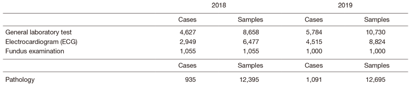
Pathology Division: All the pathologists engage in surgical pathology and research into cancer biology or development of new drugs. The number of samples examined at the department in 2018 is listed in Table1.
Clinical Laboratory Division: This division consists of eight sections; 1) general laboratory medicine, 2) hematology, 3) biochemistry and immunology, 4) physiological function testing, 5) bacteriology, 6) blood transfusion, 7) pathology and genetic testing, and 8) supporting laboratory testing in clinical studies. The numbers of tests performed in each division are listed in Table 2 and 3.
The total number of tests performed in the DPCL in 2018 increased about 7% compared with the previous year.
Since October 2018, we have converted all the tests performed in sections 1, 2 and 3 from outside laboratory control into in-house management. Thus, we centralized our management into one organizational organ.
Table 1. Number of pathology and cytology samples examined in the Pathology Division in 2019
Table 2. Number of laboratory tests examined in the Clinical Laboratory Division in 2018 & 2019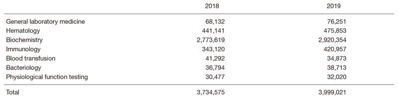
Research activities
All of the pathologists were involved in research activities at the Exploratory Oncology Research and Clinical Trial Center (EPOC). We made strived to keep good quality formalin fixed paraffin embedded specimens and facilitated numerous research projects. Especially, we contributed to SCRUM-Japan (Cancer Genome Screening Project for Individualized Medicine in Japan) by using surgical specimens.
Clinical trials
The supporting laboratory testing in clinical studies, coordinated with pathology and physiological function testing, reinforces quality control and quality assurance for clinical testing performed in clinical trials at the NCCHE. Practically the Clinical Laboratory Division participated in all of the clinical trials operated at the NCCHE by providing laboratory data.
Education
The staff of the Clinical Laboratory Division regularly conduct study sessions or lecture meetings in order to improve their expertise and testing technique based on the requirements of ISO 15189.
Clinicopathological conferences are held regularly with each clinical department/section. In the Pathology Division, conference-style training sessions are open weekly for the residents.
Future prospects
Pathological diagnosis and laboratory tests play a fundamental role not just in routine hospital work but also in medical research.
We have acquired international standard ISO15189 authorization, and will push on with pathological diagnosis, laboratory tests, and clinical studies.
In 2022, we will acquire CAP (College of American Pathologists) certification in order to form the basis for research and development at East Hospital and to be actively involved in the development of new therapies.
The DPCL will be continuously involved in investing in new diagnostic technologies, developing new drugs and conducting translational /clinical research in the NCCHE.
List of papers published in 2019
Journal
1. Sekihara K, Aokage K, Miyoshi T, Tane K, Ishii G, Tsuboi M. Perioperative pirfenidone treatment as prophylaxis against acute exacerbation of idiopathic pulmonary fibrosis: a single-center analysis. Surg Today, 50:905-911, 2020
2. Hatogai K, Fujii S, Kitano S, Kojima T, Daiko H, Yoshino T, Ohtsu A, Takiguchi Y, Doi T, Ochiai A. Relationship between the immune microenvironment of different locations in a primary tumour and clinical outcomes of oesophageal squamous cell carcinoma. Br J Cancer, 122:413-420, 2020
3. Kakinuma R, Muramatsu Y, Asamura H, Watanabe SI, Kusumoto M, Tsuchida T, Kaneko M, Tsuta K, Maeshima AM, Ishii G, Nagai K, Yamaji T, Matsuda T, Moriyama N. Low-dose CT lung cancer screening in never-smokers and smokers: results of an eight-year observational study. Transl Lung Cancer Res, 9:10-22, 2020
4. Takashima K, Fujii S, Komatsuzaki R, Komatsu M, Takahashi M, Kojima T, Daiko H, Minashi K, Chiwaki F, Muto M, Sasaki H, Yano T. CD24 and CK4 are upregulated by SIM2, and are predictive biomarkers for chemoradiotherapy and surgery in esophageal cancer. Int J Oncol, 56:835-847, 2020
5. Kubota Y, Kawazoe A, Sasaki A, Mishima S, Sawada K, Nakamura Y, Kotani D, Kuboki Y, Taniguchi H, Kojima T, Doi T, Yoshino T, Ishii G, Kuwata T, Shitara K. The Impact of Molecular Subtype on Efficacy of Chemotherapy and Checkpoint Inhibition in Advanced Gastric Cancer. Clin Cancer Res, 26:3784-3790, 2020
6. Sugiyama E, Togashi Y, Takeuchi Y, Shinya S, Tada Y, Kataoka K, Tane K, Sato E, Ishii G, Goto K, Shintani Y, Okumura M, Tsuboi M, Nishikawa H. Blockade of EGFR improves responsiveness to PD-1 blockade in EGFR-mutated non-small cell lung cancer. Sci Immunol, 5:eaav3937. doi: 10.1126/sciimmunol.aav3937, 2020
7. Maeda M, Takeshima H, Iida N, Hattori N, Yamashita S, Moro H, Yasukawa Y, Nishiyama K, Hashimoto T, Sekine S, Ishii G, Ochiai A, Fukagawa T, Katai H, Sakai Y, Ushijima T. Cancer cell niche factors secreted from cancer-associated fibroblast by loss of H3K27me3. Gut, 69:243-251, 2020
8. Yamamoto Y, Kadota T, Yoda Y, Hori K, Hatogai K, Kojima T, Fujii S, Akimoto T, Yano T. Review of early endoscopic findings in patients with local recurrence after definitive chemoradiotherapy for esophageal squamous cell carcinoma. Esophagus, 2020
9. Hiyama T, Kuno H, Nagaki T, Sekiya K, Oda S, Fujii S, Hayashi R, Kobayashi T. Extra-nodal extension in head and neck cancer: how radiologists can help staging and treatment planning. Jpn J Radiol, 38:489-506, 2020
10. Fujii S, Lam AK. Macroscopic Assessment and Sampling of Endoscopic Resection Specimens for Squamous Epithelial Malignancies with Superficial Involvement of Esophagus. Methods Mol Biol, 2129:63-81, 2020
11. Shinmura K, Ikematsu H, Kojima M, Nakamura H, Osera S, Yoda Y, Hori K, Oono Y, Ochiai A, Yano T. Safety of endoscopic procedures with monopolar versus bipolar instruments in an ex vivo porcine model. BMC Gastroenterol, 20:27, 2020
12. Okuyama H, Ikeda M, Okusaka T, Furukawa M, Ohkawa S, Hosokawa A, Kojima Y, Hara H, Murohisa G, Shioji K, Asagi A, Mizuno N, Kojima M, Yamanaka T, Furuse J. A phase II trial of everolimus in patients with advanced pancreatic neuroendocrine carcinoma refractory or intolerant to platinum-containing chemotherapy (NECTOR trial). Neuroendocrinology, 2020
13. Imaizumi K, Suzuki T, Kojima M, Shimomura M, Sakuyama N, Tsukada Y, Sasaki T, Nishizawa Y, Taketomi A, Ito M, Nakatsura T. Ki67 expression and localization of T cells after neoadjuvant therapies as reliable predictive markers in rectal cancer. Cancer Sci, 111:23-35, 2020
14. Kojima M, Yamauchi C, Oyamada S, Hojo T, Iwase S, Naito A, Yamano K, Takahashi S, Ochiai A. Assessment of Upper Limb Physiological Features in Patients with Lymphedema After Breast Surgery Using Multiple Instruments. Lymphat Res Biol, 18:239-246, 2020
15. Ogawa K, Kobayashi N, Miyagawa F, Nakai T, Fukumoto T, Mitsui Y, Arai E, Asada H. Case of nodal nevus with melanocytic cell aggregates in the lymphatic hilum: A potential diagnostic pitfall that requires differentiation from metastatic melanoma of the lymph node. J Dermatol, 47:e242-e244, 2020
16. Sugita S, Kinoshita T, Kuwata T, Tokunaga M, Kaito A, Watanabe M, Tonouchi A, Sato R, Nagino M. Long-term oncological outcomes of laparoscopic versus open transhiatal resection for patients with Siewert type II adenocarcinoma of the esophagogastric junction. Surg Endosc, 2020
17. Miyashita T, Neri S, Hashimoto H, Akutsu A, Sugano M, Fujii S, Ochiai A, Ishii G. Fibroblasts-dependent invasion of podoplanin-positive cancer stem cells in squamous cell carcinoma. J Cell Physiol, 235:7251-7260, 2020
18. Suzuki J, Kojima M, Aokage K, Sakai T, Nakamura H, Ohara Y, Tane K, Miyoshi T, Sugano M, Fujii S, Kuwata T, Ochiai A, Ito M, Suzuki K, Tsuboi M, Ishii G. Clinicopathological characteristics associated with necrosis in pulmonary metastases from colorectal cancer. Virchows Arch, 474:569-575, 2019
19. Oki T, Hishida T, Yoshida J, Goto M, Sekihara K, Miyoshi T, Aokage K, Ishii G, Tsuboi M. Survival and prognostic factors after pulmonary metastasectomy of head and neck cancer: what are the clinically informative prognostic indicators? Eur J Cardiothorac Surg, 55:942-947, 2019
20. Naito T, Umemura S, Nakamura H, Zenke Y, Udagawa H, Kirita K, Matsumoto S, Yoh K, Niho S, Motoi N, Aokage K, Tsuboi M, Ishii G, Goto K. Successful treatment with nivolumab for SMARCA4-deficient non-small cell lung carcinoma with a high tumor mutation burden: A case report. Thorac Cancer, 10:1285-1288, 2019
21. Omori T, Aokage K, Nakamura H, Katsumata S, Miyoshi T, Sugano M, Kojima M, Fujii S, Kuwata T, Ochiai A, Ikeda N, Tsuboi M, Ishii G. Growth patterns of small peripheral squamous cell carcinoma of the lung and their impacts on pathological and biological characteristics of tumor cells. J Cancer Res Clin Oncol, 145:1773-1783, 2019
22. Miyoshi T, Aokage K, Katsumata S, Tane K, Ishii G, Tsuboi M. Ground-Glass Opacity Is a Strong Prognosticator for Pathologic Stage IA Lung Adenocarcinoma. Ann Thorac Surg, 108:249-255, 2019
23. Nakamura H, Sugano M, Miyashita T, Hashimoto H, Ochiai A, Suzuki K, Tsuboi M, Ishii G. Organoid culture containing cancer cells and stromal cells reveals that podoplanin-positive cancer-associated fibroblasts enhance proliferation of lung cancer cells. Lung Cancer, 134:100-107, 2019
24. Katsumata S, Aokage K, Ishii G, Nakasone S, Sakai T, Okada S, Miyoshi T, Tane K, Tsuboi M. Prognostic Impact of the Number of Metastatic Lymph Nodes on the Eighth Edition of the TNM Classification of NSCLC. J Thorac Oncol, 14:1408-1418, 2019
25. Suzuki E, Yamazaki S, Naito T, Hashimoto H, Okubo S, Udagawa H, Goto K, Tsuboi M, Ochiai A, Ishii G. Secretion of high amounts of hepatocyte growth factor is a characteristic feature of cancer-associated fibroblasts with EGFR-TKI resistance-promoting phenotype: A study of 18 cases of cancer-associated fibroblasts. Pathol Int, 69:472-480, 2019
26. Ota T, Niho S, Kirita K, Ishii G, Tsuboi M, Goto K. Impressive response to nivolumab of non-small-cell lung cancer containing sarcomatoid components. Respirol Case Rep, 7:e00477, 2019
27. Oki T, Aokage K, Ueda T, Sugano M, Tane K, Miyoshi T, Kojima M, Fujii S, Kuwata T, Ochiai A, Funai K, Tsuboi M, Ishii G. Proportion of goblet cell is associated with malignant potential in invasive mucinous adenocarcinoma of the lung. Pathol Int, 69:526-535, 2019
28. Ichikawa T, Aokage K, Miyoshi T, Tane K, Suzuki K, Makinoshima H, Tsuboi M, Ishii G. Correlation between maximum standardized uptake values on FDG-PET and microenvironmental factors in patients with clinical stage IA radiologic pure-solid lung adenocarcinoma. Lung Cancer, 136:57-64, 2019
29. Hisamitsu S, Miyashita T, Hashimoto H, Neri S, Sugano M, Nakamura H, Yamazaki S, Ochiai A, Goto K, Tsuboi M, Ishii G. Interaction between cancer cells and cancer-associated fibroblasts after cisplatin treatment promotes cancer cell regrowth. Hum Cell, 32:453-464, 2019
30. Naito T, Udagawa H, Umemura S, Sakai T, Zenke Y, Kirita K, Matsumoto S, Yoh K, Niho S, Tsuboi M, Ishii G, Goto K. Non-small cell lung cancer with loss of expression of the SWI/SNF complex is associated with aggressive clinicopathological features, PD-L1-positive status, and high tumor mutation burden. Lung Cancer, 138:35-42, 2019
31. Sunami K, Ichikawa H, Kubo T, Kato M, Fujiwara Y, Shimomura A, Koyama T, Kakishima H, Kitami M, Matsushita H, Furukawa E, Narushima D, Nagai M, Taniguchi H, Motoi N, Sekine S, Maeshima A, Mori T, Watanabe R, Yoshida M, Yoshida A, Yoshida H, Satomi K, Sukeda A, Hashimoto T, Shimizu T, Iwasa S, Yonemori K, Kato K, Morizane C, Ogawa C, Tanabe N, Sugano K, Hiraoka N, Tamura K, Yoshida T, Fujiwara Y, Ochiai A, Yamamoto N, Kohno T. Feasibility and utility of a panel testing for 114 cancer-associated genes in a clinical setting: A hospital-based study. Cancer Sci, 110:1480-1490, 2019
32. Okada N, Daiko H, Kanamori J, Sato A, Horikiri Y, Sato T, Fujiwara H, Tomioka T, Fujita T, Kojima T, Fujii S. Impact of pathologically assessing extranodal extension in the thoracic field on the prognosis of esophageal squamous cell carcinoma. Surgery, 165:1203-1210, 2019
33. Naito T, Udagawa H, Sato J, Horinouchi H, Murakami S, Goto Y, Kanda S, Fujiwara Y, Yamamoto N, Zenke Y, Kirita K, Matsumoto S, Yoh K, Niho S, Motoi N, Ohe Y, Ishii G, Goto K. A Minimum Of 100 Tumor Cells in a Single Biopsy Sample Is Required to Assess Programmed Cell Death Ligand 1 Expression in Predicting Patient Response to Nivolumab Treatment in Nonsquamous Non-Small Cell Lung Carcinoma. J Thorac Oncol, 14:1818-1827, 2019
34. Ota T, Kirita K, Matsuzawa R, Udagawa H, Matsumoto S, Yoh K, Niho S, Ishii G, Goto K. Validity of using immunohistochemistry to predict treatment outcome in patients with non-small cell lung cancer not otherwise specified. J Cancer Res Clin Oncol, 145:2495-2506, 2019
35. Ghani FI, Dendo K, Watanabe R, Yamada K, Yoshimatsu Y, Yugawa T, Nakahara T, Tanaka K, Yoshida H, Yoshida M, Ishikawa M, Goshima N, Kato T, Kiyono T. An Ex-Vivo Culture System of Ovarian Cancer Faithfully Recapitulating the Pathological Features of Primary Tumors. Cells, 8:644, 2019
36. Ohashi-Nakatani K, Shibuki Y, Fujima M, Watanabe R, Yoshida A, Yoshida H, Matsumoto Y, Tsuchida T, Watanabe SI, Motoi N. Primary pulmonary meningioma: A rare case report of aspiration cytological features and immunohistochemical assessment. Diagn Cytopathol, 47:330-333, 2019
37. Sasaki A, Nakamura Y, Mishima S, Kawazoe A, Kuboki Y, Bando H, Kojima T, Doi T, Ohtsu A, Yoshino T, Kuwata T, Akimoto T, Shitara K. Predictive factors for hyperprogressive disease during nivolumab as anti-PD1 treatment in patients with advanced gastric cancer. Gastric Cancer, 22:793-802, 2019
38. Nakamura Y, Sawada K, Fujii S, Yoshino T. HER2-targeted therapy should be shifted towards an earlier line for patients with anti-EGFR-therapy na?ve, HER2-amplified metastatic colorectal cancer. ESMO Open, 4:e000530, 2019
39. Kaito A, Kuwata T, Tokunaga M, Shitara K, Sato R, Akimoto T, Kinoshita T. HER2 heterogeneity is a poor prognosticator for HER2-positive gastric cancer. World J Clin Cases, 7:1964-1977, 2019
40. Fujii S, Yoshino T, Yamazaki K, Muro K, Yamaguchi K, Nishina T, Yuki S, Shinozaki E, Shitara K, Bando H, Mimaki S, Nakai C, Matsushima K, Suzuki Y, Akagi K, Yamanaka T, Nomura S, Esumi H, Sugiyama M, Nishida N, Mizokami M, Koh Y, Abe Y, Ohtsu A, Tsuchihara K. Histopathological factors affecting the extraction of high quality genomic DNA from tissue sections for next-generation sequencing. Biomed Rep, 11:171-180, 2019
41. Okubo S, Kojima M, Matsuda Y, Hioki M, Shimizu Y, Toyama H, Morinaga S, Gotohda N, Uesaka K, Ishii G, Mino-Kenudson M, Takahashi S. Area of residual tumor (ART) can predict prognosis after post neoadjuvant therapy resection for pancreatic ductal adenocarcinoma. Sci Rep, 9:17145, 2019
42. Maishi N, Kikuchi H, Sato M, Nagao-Kitamoto H, Annan DA, Baba S, Hojo T, Yanagiya M, Ohba Y, Ishii G, Masutomi K, Shinohara N, Hida Y, Hida K. Development of Immortalized Human Tumor Endothelial Cells from Renal Cancer. Int J Mol Sci, 20:4595, 2019
43. Sakai T, Miyoshi T, Umemura S, Suzuki J, Nakasone S, Okada S, Tane K, Aokage K, Goto K, Motoi N, Ishii G, Tsuboi M. Large pulmonary sclerosing pneumocytoma with massive necrosis and vascular invasion: a case report. Oxf Med Case Reports, 2019:omz066, 2019
44. Martin JD, Panagi M, Wang C, Khan TT, Martin MR, Voutouri C, Toh K, Papageorgis P, Mpekris F, Polydorou C, Ishii G, Takahashi S, Gotohda N, Suzuki T, Wilhelm ME, Melo VA, Quader S, Norimatsu J, Lanning RM, Kojima M, Stuber MD, Stylianopoulos T, Kataoka K, Cabral H. Dexamethasone Increases Cisplatin-Loaded Nanocarrier Delivery and Efficacy in Metastatic Breast Cancer by Normalizing the Tumor Microenvironment. ACS Nano, 13:6396-6408, 2019
45. Oono Y, Shinmura K, Hori K, Yoda Y, Ishii G, Ikematsu H, Yano T. Endoscopic submucosal resection using a ligation device without injection for duodenal neuroendocrine tumors. Surg Endosc, 33:2008-2014, 2019
46. Sukeda A, Nakamura Y, Nishida Y, Kojima M, Gotohda N, Akimoto T, Ochiai A. Expression of Monocarboxylate Transporter 1 Is Associated With Better Prognosis and Reduced Nodal Metastasis in Pancreatic Ductal Adenocarcinoma. Pancreas, 48:1102-1110, 2019
47. Hashimoto T, Ogawa R, Yoshida H, Taniguchi H, Kojima M, Saito Y, Sekine S. EIF3E-RSPO2 and PIEZO1-RSPO2 fusions in colorectal traditional serrated adenoma. Histopathology, 75:266-273, 2019
48. Matsunaga R, Kojima M, Nishizawa Y, Yokota M, Hasegawa H, Saito N, Ito M, Ochiai A. The utility of longitudinal slicing method for rectal specimen: pathological analysis of circumferential resection margin of intersphincteric resection for low-lying rectal cancer. Pathol Int, 69:272-281, 2019
49. Nakai T, Okamoto Y. A megakaryocyte in a peripheral blood smear. Int J Hematol, 110:647, 2019
50. Tonouchi A, Sugano M, Tokunaga M, Sugita S, Watanabe M, Sato R, Kaito A, Akimoto T, Ochiai A, Kinoshita T, Kuwata T. Extra-perigastric Extranodal Metastasis is a Significant Prognostic Factor in Node-Positive Gastric Cancer. World J Surg, 43:2499-2505, 2019
51. Kawai H, Sugano M, Nakano N, Muratani M, Sakashita S, Goto Y, Noguchi M. A case of invasive mucinous adenocarcinoma of the lung showing stepwise progression at the primary site. Lung Cancer, 136:94-97, 2019
52. Kuwata T, Yanagihara K, Iino Y, Komatsu T, Ochiai A, Sekine S, Taniguchi H, Katai H, Kinoshita T, Ohtsu A. Establishment of Novel Gastric Cancer Patient-Derived Xenografts and Cell Lines: Pathological Comparison between Primary Tumor, Patient-Derived, and Cell-Line Derived Xenografts. Cells, 8:585, 2019
53. Nishihara K, Oono Y, Kuwata T, Ikematsu H, Yano T. Depressed gastric-type adenoma in nonatrophic gastric mucosa without Helicobacter pylori infection. Endoscopy, 51:E138-E140, 2019
Book
1. WHO classification of Tumours (ed), Breast Tumours, WHO Classification of Tumours, 5th Edition, Volume 2, Lyon, IARC, 2019
2. WHO classification of Tumours (ed), Digestive System Tumours WHO Classification of Tumours, 5th Edition, Lyon, IARC, 2019