Annual Report 2020
Department of Diagnostic Radiology
Tatsushi Kobayashi, Hirofumi Kuno, Yasunori Arai, Kaoru Shimada, Takashi Hiyama, Shioto Oda, Rakuhei Nakama, Kotaro Hosoi
Introduction
The Department of Diagnostic Radiology is committed to improving health through excellence in image-oriented patient care and research. Our department performs approximately 120,000 inpatient and outpatient examinations annually. Our department also conducts clinical scientific research and basic scientific studies, with the results translated directly into better patient care.
The Team and What We Do
Our department has four multi-slice CT scanners including one ultra-high-resolution CT, two area-detector CT scanners and one dual-source CT, two 3T MRI systems, one interventional radiology (IR) CT system, one Multi-axis c-arm CT system, two gamma cameras with the capacity for single photon emission CT (SPECT), two digital radiographic (DR) systems for fluoroscopy, two mammographies (MMG) machines, and four computed radiographic (CR) systems. Our IR-CT systems use digital subtraction angiography with multi-detector computerized tomography (MDCT). One is equipped with a 320 multi-slice CT. A positron emission tomography (PET) scanner and baby cyclotron have been installed, and tumor imaging using 18F-FDG (fluorodeoxyglucose) has been performed. These all-digital image systems enhance the efficacy of routine examinations.
This department has eight consulting radiologists and 22 technologists. As part of our routine activities, every effort is made to produce an integrated report covering almost all examinations, such as MMG, contrast radiological procedures, CT, MRI, RI, PET, angiography and IR, mainly transarterial chemoembolization (TACE).
The number of cases examined in 2020 is shown in Table 1 and Table 2. Several conferences are routinely held at our department including pre- and postoperative conferences. Our department also contributes to deciding the treatment strategy through image presentations at the weekly tumor board conference (especially, the hepatobiliary-pancreatic and head-neck regions).
Table 1. Number of examinations in 2020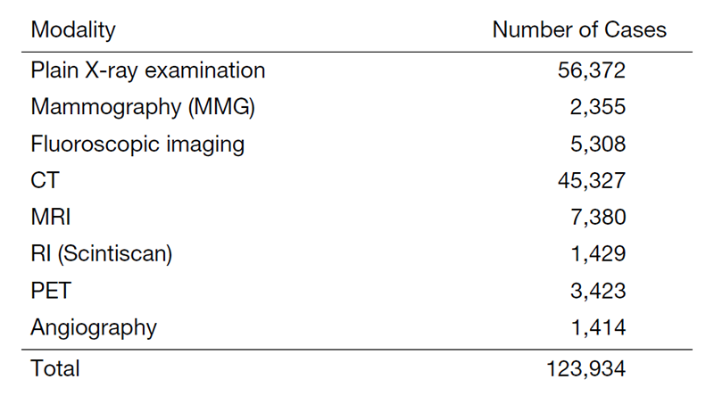
Table 2. Number of Interventional Radiology Procedures in 2020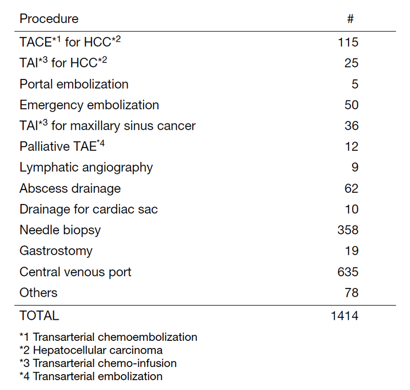
Research activities
The research activities of the Department of Diagnostic Radiology focus on diagnostic imaging and IR. These activities consist of: 1) Development of new CT/MR imaging techniques and 2) Texture analysis and radiomics research. Our department also conducts clinical scientific research and basic scientific studies, the results of which are translated directly into better patient care.
(1) Development of new CT/MR imaging techniques
A major focus of our department is on the development of new imaging techniques using advanced CT/MR systems including dual-energy CT (DECT) and area-detector CT (ADCT) for cancer patients.
DECT has the potential to improve the detection of pathologies and increase diagnostic confidence in the evaluation of a variety of cancers by using X-ray energy-dependent absorption behaviors of various materials. DECT allows for material decomposition so that iodine can be differentiated from soft tissue, and it can potentially provide additional further "contrast resolution" to standard contrast-enhanced CT images. We work with dual-source CT and ADCT systems, focusing mostly on head and neck and pancreatic cancer imaging, using iodine overlay images that are generated using three-material decomposition algorithms.
We also work on the new imaging technique "bone subtraction iodine (BSI) imaging" that uses 320-row ADCT scanning. This additional method can include subtracting unenhanced CT from contrast-enhanced CT using subtraction software recently employed in oncology imaging. This technique reduces spatial mismatch using volume scanning with wide ADCT and a high-resolution deformable registration algorithm, and enables the identification of contrast enhancement in the bone marrow. BSI imaging using 320-row ADCT scanning is therefore expected to be useful for detecting bone invasion, such as in the skull base/mandible, and accurately assessing the extent of bone and soft tissue invasion by cancer cells. We are working on several validation studies such as the evaluation of bone/soft tissue invasion in patients with nasopharyngeal carcinoma or oral cavity cancer.
(2) Texture analysis and radiomics research
Our department also focuses on developing new techniques to determine diagnoses and to predict prognoses, treatment response, and outcomes from images and other associated data using texture analysis techniques and "radiomics". Image texture is defined as a complex visual pattern within an image consisting of simpler sub-patterns with characteristic features, and texture analysis allows the mathematical detection of the subtle spatial arrangement of the gray levels among image pixels. Furthermore, "radiomics" extends traditional imaging consultation to include a deeper analysis of these medical images from various imaging modalities (e.g., CT, PET, and MRI) and refers to the extraction and analysis of a large number of advanced quantitative imaging texture features with high throughput. These radiomics data will have an impact on personalized medicine, where treatment can be tailored towards patient-specific needs. One of our main areas of interest is, therefore, connecting tumor-specific radiomic features with their clinical information, including treatment outcome. Ultimately, we aim to translate these developments into clinical applications and decision support systems using machine learning algorithms. We primarily work with cross-sectional images, including CT and MRI, and specialize in cancer imaging, focusing mostly on head and neck and pancreatic cancers.
Clinical trials
We are conducting a multicenter clinical trial, "The single-armed confirmatory trial for immediate effectivity and safety of palliative arterial embolization for painful bone metastases. (JIVROSG/J-SUPPORT 1903)", as a representative facility, which is scheduled to start later this year. The purpose of this trial is to verify the safety and immediate effect of transarterial embolization as a palliative treatment for painful bone metastases and to establish it as a standard treatment. This research is funded by the Japan Agency for Medical Research and Development (AMED).
List of papers published in 2020
Journal
1. Sekihara K, Aokage K, Hiyama T, Oiwa H, Miyoshi T, Tane K, Ishii G, Tsuboi M. Prognostic impact of home oxygen therapy on patients with resected non-small-cell lung cancer with interstitial lung disease. Surg Today, 51:1036-1043, 2021
2. Hiyama T, Kuno H, Sekiya K, Oda S, Kobayashi T. Imaging of Malignant Minor Salivary Gland Tumors of the Head and Neck. Radiographics, 41:175-191, 2021
3. Yasuta S, Kobayashi T, Aizawa H, Takahashi S, Ikeda M, Konishi M, Kojima M, Kuno H, Uesaka K, Morinaga S, Miyamoto A, Toyama H, Takakura N, Sugimachi K, Takayama W. Relationship between surgical R0 resectability and findings of peripancreatic vascular invasion on CT imaging after neoadjuvant S-1 and concurrent radiotherapy in patients with borderline resectable pancreatic cancer. BMC Cancer, 20:1184, 2020
4. Tomita H, Kuno H, Sekiya K, Otani K, Sakai O, Li B, Hiyama T, Nomura K, Mimura H, Kobayashi T. Quantitative Assessment of Thyroid Nodules Using Dual-Energy Computed Tomography: Iodine Concentration Measurement and Multiparametric Texture Analysis for Differentiating between Malignant and Benign Lesions. Int J Endocrinol, 2020:5484671, 2020
Book
1. Kuno H. Diagnostic Imaging of Laryngeal and Hypopharyngeal Cancers. In: Diagnostic Imaging in Head and Neck Cancer, Singapore, Springer Singapore, pp 75-111, 2020
2. Takashi H. Diagnostic Imaging of Oral Cavity Cancer. In: Hiroya O (ed), Diagnostic Imaging in Head and Neck Cancer, Singapore, Springer Singapore, pp 51-73, 2020