Annual Report 2021
Department of Radiological Technology
Yoshihisa Muramatsu, Kazutoshi Yokoyama, Yuichi Nagai, Tetsuya Aoyama, Masashi Iseya, Takaki Ariji, Akira Inagaki, Naotaka Yamazawa, Atsuko Takada, Satoe Kito, Masufumi Shinozaki, Tsunemichi Akita, Hajime Ohyoshi, Keiichi Nomura, Hiroyuki Ota, Shuhei Ohashi, Toshiya Rachi, Asami Tanaka, Yuki Tanaka, Shun Aoyagi, Kaori Yanagisawa, Toshiyuki Shibuya, Kota Hirotaki, Kazuto Kano, Taku Tochinai, Chikara Mano, Kazuya Mizuguchi, Takara Watanabe, Hikari Inagawa, Moeka Mizuguchi, Naoki Yamashita, Takashi Someya, Hiroaki Sagara, Yumika Hirai, Taknori Matsuoka, Kosei Hamagashira, Ken Hirayama, Amon Ohsawa, Nene Hashizume, Haruki Togo, Saeko Mochinaga, Yori Takaki, Yuya Fujimoto, Yuki Mizunuma, Risa Ono, Kento Yokota, Kazusa Honda, Yuta Suzuki, Mai Umehara, Kurumi Aoki, Chiaki Tagami, Mitsue Nihei, Motoe Kiyama, Hiroe Ishii
Introduction
The overall number of cases increased by 105% from the previous year (Table 1). The breakdown shows a large increase in the number of IVR examinations and radiotherapy, especially IMRT.
Table 1. Transition in the number of radiological examinations
and radiation therapies by year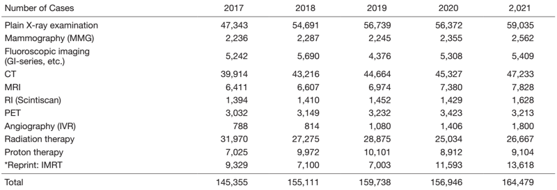
The Team and What We Do
A new remote afterloading system (RALS) for radioactive sources for brachytherapy was installed, and a C-arm fluoroscopy system, a dental panoramic system, a mammography system, an X-TV system, and an IVR-CT system were upgraded. Use of Lutetium (177Lu) oxodotreotide (Lutathera®) was also started.
In research activities, joint research agreements were signed with six companies, and translational research on clinical technologies continued to produce results. The main areas of research are next-generation CT systems, 3D imaging with remote access, AI-based image QA systems, radiation dose management systems in the RI section, and the potential benefits of adaptive radiation therapy.
Research activities
Organ doses were obtained from dose estimates of chest-abdomen-pelvis CT examinations in pediatric patients, and their correlations with dose indices were examined. Both showed a high linear correlation and could be easily estimated using linear regression.
In PET examinations, the effectiveness of the operation to determine the dose by the subject's body weight specified in the DRLs (Diagnostic Reference Levels) 2020 was evaluated. This operation was shown to contribute not only to the reduction of effective doses but also to the improvement of image quality in standard body types.
The effectiveness of the method of converting the dose distribution into energy fluence and reproducing it with a multi-leaf collimator was demonstrated on the basis of clinical cases in the replanning from an O-ring type to a C-type linear accelerator, which has a different mechanical structure.
A system was developed to detect anatomical changes in adaptive radiation therapy using water equivalent thickness calculated based on 2D X-ray images as an indicator. This system can observe daily anatomical changes without requiring high radiation exposure or inefficient work.
A useful index was devised to quantify and compare the magnitude of textural noise found in CT images reconstructed by iterative reconstruction (IR) and deep learning reconstruction (DLR) algorithms.
Education
The Human Resource Development Center is taking the lead in job rotation to maintain and improve the functions of each department.
In the Radiological Technology Department, in addition to the Clinical Coordinator Office, Medical Information Department, and Ethics Review Office, a new assignment to the Medical Affairs Management Section was implemented, as well as a secondment to the Japan Agency for Medical Research and Development (AMED).
Future Prospects
As the workload continues to increase, we will be welcoming our first class of residents in FY2022. We will be expected to achieve more visible results and accomplishments as medical treatment, research, and education become balanced.
List of papers published in 2021
Journal
1. Hirotaki K, Moriya S, Tachibana H, Sakae T. Detection of anatomical changes using two-dimensional x-ray images for head and neck adaptive radiotherapy. Medical physics, 49:3288-3297, 2022
2. Rachi T, Parshuram RV, Tanaka Y, Togo H. Examination of conversion method of dose distribution of lung cancer IMRT using fluence reversible calculation function in O-ring type linac and C-type linac. Physical and engineering sciences in medicine, 2022
3. Fujii K, Nomura K, Imai K, Muramatsu Y, Tsushima S, Ota H. Evaluation of Apparent Noise on CT Images Using Moving Average Filters. Journal of digital imaging, 35:77-85, 2022
4. Raturi VP, Motegi A, Zenda S, Nakamura N, Hojo H, Kageyama SI, Okumura M, Rachi T, Ohyoshi H, Tachibana H, Motegi K, Ariji T, Nakamura M, Hirano Y, Hirata H, Akimoto T. Comparison of a Hybrid IMRT/VMAT technique with non-coplanar VMAT and non-coplanar IMRT for unresectable olfactory neuroblastoma using the RayStation treatment planning system-EUD, NTCP and planning study. Journal of radiation research, 62:540-548, 2021
5. Sagara H, Inoue K, Yaku H, Ohsawa A, Someya T, Yanagisawa K, Ohashi S, Ishigaki R, Wakabayashi M, Muramatsu Y, Fujii H. Optimization of injection dose in 18F-FDG PET/CT based on the 2020 national diagnostic reference levels for nuclear medicine in Japan. Annals of nuclear medicine, 35:1177-1186, 2021
6. Fujii K, Nomura K, Muramatsu Y, Ota H. PATIENT-SPECIFIC ORGAN DOSE EVALUATION BASED ON MONTE CARLO SIMULATION AND DOSE METRICS IN PAEDIATRIC CHEST-ABDOMEN-PELVIS CT EXAMINATIONS. Radiation protection dosimetry, 197:46-53, 2021