
Home > Organization > Division of Functional Imaging(Kashiwa) > Research Summary > Development of unique imaging probes
Development of unique imaging probes
We are engaged in the development of molecular imaging probes that can visualize information expressed in tumor lesions or that show high affinity to tumor foci.
Development of imaging probes that can visualize hypoxic areas in tumors
Visualization of hypoxic areas inside tumor lesions would be useful to optimize the treatment of cancer because these areas are usually resistant to radiotherapy and/or chemotherapy. We have proposed a series of unique Tc-99m labeled molecular probes that can stay in the hypoxic regions inside tumors based on the reductive reaction of nitro-imidazole compounds.
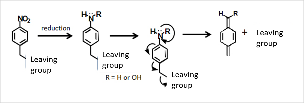
- Kimura S, Umeda IO, Moriyama N, Fujii H. Synthesis and evaluation of a novel 99mTc-labeled bioreductive probe for tumor hypoxia imaging. Bioorg Med Chem Lett, 21:7359-7362, 2011 [PubMed]
Development of iron oxide nanoparticles that show high affinity to tumors
We have developed iron oxide nanoparticles coated by PEG-b-PVBP that are stable in the blood without coagulation in collaboration with Professor Nagasaki’s team at Tsukuba University. These nanoparticles accumulated in tumors implanted to mice with a good ratio of over 15% ID/g・tumor, which was higher than that of conventional iron oxide nanoparticles. These nanoparticles are expected to be applicable to clinical practice as MR contrast media or sensitizer for thermal therapy.
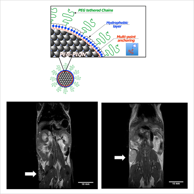
- Reprinted from Colloids Surf B Biointerfaces, Ujiie K, et al., Preparation of highly dispersible and tumor-accumulative, iron oxide nanoparticles Multi-point anchoring of PEG-b-poly(4-vinylbenzylphosphonate) improves performance significantly, 771-778, Copyright (2011), with permission from Elsevier.
- Ujiie K, Kanayama N, Asai K, Kishimoto M, Ohara Y, Akashi Y, Yamada K, Hashimoto S, Oda T, Ohkohchi N, Yanagihara H, Kita E, Yamaguchi M, Fujii H, Nagasaki Y. Preparation of highly dispersible and tumor-accumulative, iron oxide nanoparticles Multi-point anchoring of PEG-b-poly(4-vinylbenzylphosphonate) improves performance significantly. Colloids Surf B Biointerfaces, 88:771-778, 2011 [PubMed]
Integrin-avid molecular probes to detect minimal pancreatic tumors
Pancreatic cancer, which is one of the most intractable tumors, can be cured only when it is detected at the early stage and the whole of the tumor is completely resected. We are interested in the abundant expression of integrin in the pancreatic cancer, and we applied 111In-DOTA-c(RGDfK), which is a molecular probe that shows strong affinity to integrin, for the detection of pancreatic tumors. This trial was successful and minimal pancreatic lesions could be visualized by in vivo SPECT/CT images.
Figure legend: The coronal SPECT image of the hamster one hour after the injection of 111In-DOTA-c(RGDfK) (left) and the photo of dissected abdomen of the hamster.
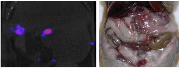
This research was originally published in JNM. Yoshimoto M, et al. In vivo SPECT Imaging with 111In-DOTA-c(RGDfK) to detect early pancreatic cancer in a hamster pancreatic carcinogenesis model. J Nucl Med. 2012; 53: 765-771. © by the Society of Nuclear Medicine and Molecular Imaging, Inc.
- Yoshimoto M, Hayakawa T, Mutoh M, Imai T, Tsuda K, Kimura S, Umeda IO, Fujii H, Wakabayashi K. In vivo SPECT Imaging with 111In-DOTA-c(RGDfK) to detect early pancreatic cancer in a hamster pancreatic carcinogenesis model. J Nucl Med, 53:765-771, 2012[PubMed]